6ULC | pdb_00006ulc

Structure of full-length, fully glycosylated, non-modified HIV-1 gp160 bound to PG16 Fab at a nominal resolution of 4.6 Angstrom


Experimental Data Snapshot

  • Method: ELECTRON MICROSCOPY
  • Resolution: 4.60 Å
  • Aggregation State: PARTICLE 
  • Reconstruction Method: SINGLE PARTICLE 

wwPDB Validation 3D Report Full Report

Validation slider image for 6ULC

This is version 2.2 of the entry. See complete history

Literature

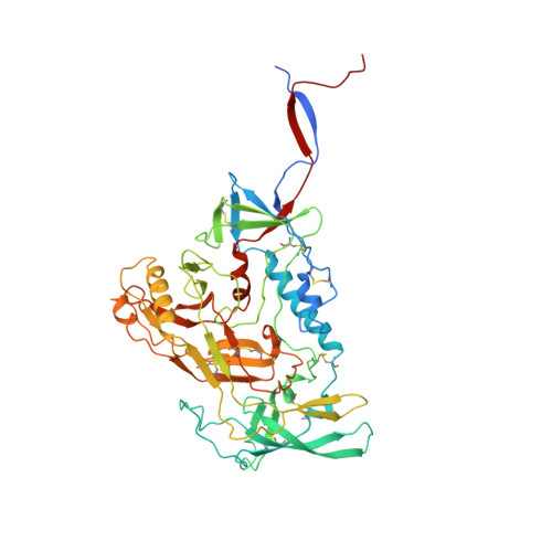
Cryo-EM Structure of Full-length HIV-1 Env Bound With the Fab of Antibody PG16.

Pan, J.Peng, H.Chen, B.Harrison, S.C.

(2020) J Mol Biology 432: 1158-1168

  • DOI: https://doi.org/10.1016/j.jmb.2019.11.028
  • Primary Citation Related Structures: 
    6PWU, 6ULC

  • PubMed Abstract: 

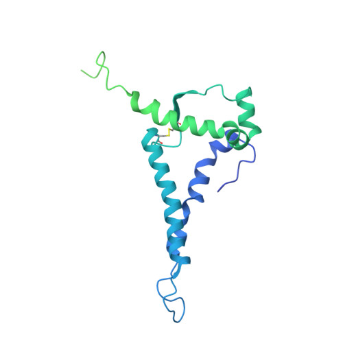
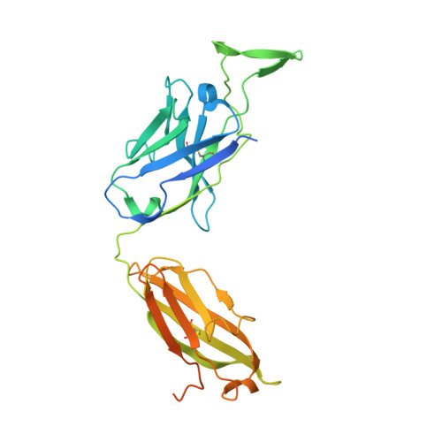
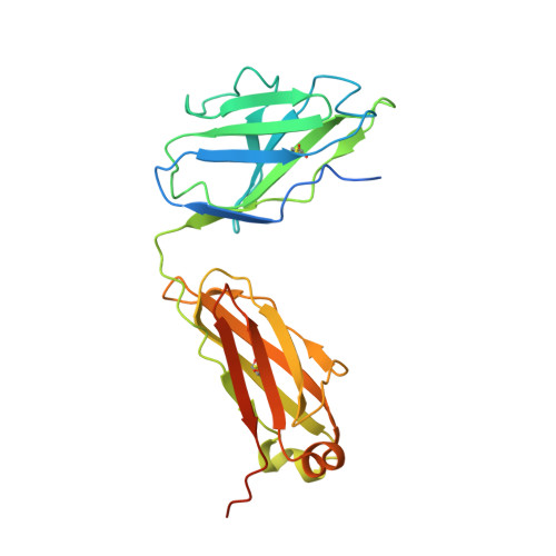
    The HIV-1 envelope protein (Env) is the target of neutralizing antibodies and the template for vaccine immunogen design. The dynamic conformational equilibrium of trimeric Env influences its antigenicity and potential immunogenicity. Antibodies that bind at the trimer apex stabilize a "closed" conformation characteristic of the most difficult to neutralize isolates. A goal of vaccine development is therefore to mimic the closed conformation in a designed immunogen. A disulfide-stabilized, trimeric Env ectodomain-the "SOSIP" construct-has many of the relevant properties; it is also particularly suitable for structure determination. Some single-molecule studies have, however, suggested that the SOSIP trimer is not a good representation of Env on the surface of a virion or an infected cell. We isolated Env (fully cleaved to gp120 and gp41) from the surface of expressing cells using tagged, apex-binding Fab PG16 and determined the structure of the PG16-Env complex by cryo-EM to an overall resolution of 4.6 Å. Placing the only purification tag on the Fab ensured that the isolated Env was continuously stabilized in its closed, native conformation. The Env structure in this complex corresponds closely to the SOSIP structures determined by both x-ray crystallography and cryo-EM. Although the membrane-interacting elements are not resolved in our reconstruction, we can make inferences about the connection between ectodomain and membrane-proximal external region (MPER) by reference to the published cryo-tomography structure of an Env "spike" and the NMR structure of the MPER-transmembrane segment. We discuss these results in view of the conflicting interpretations in the literature.


  • Organizational Affiliation
    • Laboratory of Molecular Medicine, Boston Children's Hospital, 3 Blackfan Circle, Boston, MA, 02115, USA.

Macromolecule Content 

  • Total Structure Weight: 379.95 kDa 
  • Atom Count: 19,968 
  • Modeled Residue Count: 2,301 
  • Deposited Residue Count: 3,121 
  • Unique protein chains: 4

Macromolecules

Find similar proteins by:|  3D Structure
Entity ID: 1
MoleculeChains  Sequence LengthOrganismDetailsImage
envelope glycoprotein gp120,envelope glycoprotein gp120
A, C, E
505Human immunodeficiency virus 1Mutation(s): 0 
Gene Names: env
UniProt
Find proteins for Q71014 (Human immunodeficiency virus type 1)
Explore Q71014 
Go to UniProtKB:  Q71014
Entity Groups
Sequence Clusters30% Identity50% Identity70% Identity90% Identity95% Identity100% Identity
UniProt GroupQ71014
Glycosylation
Glycosylation Sites: 24
Sequence Annotations
Expand
Reference Sequence
Find similar proteins by:|  3D Structure
Entity ID: 2
MoleculeChains  Sequence LengthOrganismDetailsImage
Envelope glycoprotein gp41
B, D, F
353Human immunodeficiency virus 1Mutation(s): 0 
Gene Names: env
UniProt
Find proteins for Q71014 (Human immunodeficiency virus type 1)
Explore Q71014 
Go to UniProtKB:  Q71014
Entity Groups
Sequence Clusters30% Identity50% Identity70% Identity90% Identity95% Identity100% Identity
UniProt GroupQ71014
Glycosylation
Glycosylation Sites: 3
Sequence Annotations
Expand
Reference Sequence
Find similar proteins by:|  3D Structure
Entity ID: 3
MoleculeChains  Sequence LengthOrganismDetailsImage
antibody PG16 Fab heavy chainG [auth H]297Homo sapiensMutation(s): 0 
Find similar proteins by:|  3D Structure
Entity ID: 4
MoleculeChains  Sequence LengthOrganismDetailsImage
antibody PG16 Fab light chainH [auth L]250Homo sapiensMutation(s): 0 

Oligosaccharides

Help  
Entity ID: 5
MoleculeChains Length2D Diagram GlycosylationD Interactions
beta-D-galactopyranose-(1-4)-2-acetamido-2-deoxy-beta-D-glucopyranose-(1-2)-alpha-D-mannopyranose-(1-6)-beta-D-mannopyranose-(1-4)-2-acetamido-2-deoxy-beta-D-glucopyranose-(1-4)-2-acetamido-2-deoxy-beta-D-glucopyranoseI [auth G]6N-Glycosylation
Glycosylation Resources
GlyTouCan: G08429SS
GlyCosmos: G08429SS
GlyGen: G08429SS
Entity ID: 6
MoleculeChains Length2D Diagram GlycosylationD Interactions
alpha-D-mannopyranose-(1-3)-[alpha-D-mannopyranose-(1-6)]beta-D-mannopyranose-(1-4)-2-acetamido-2-deoxy-beta-D-glucopyranose-(1-4)-2-acetamido-2-deoxy-beta-D-glucopyranoseJ [auth I],
K [auth J]
5N-Glycosylation
Glycosylation Resources
GlyTouCan: G22768VO
GlyCosmos: G22768VO
GlyGen: G22768VO
Entity ID: 8
MoleculeChains Length2D Diagram GlycosylationD Interactions
2-acetamido-2-deoxy-beta-D-glucopyranose-(1-4)-2-acetamido-2-deoxy-beta-D-glucopyranose
BA [auth b],
HA [auth h],
LA [auth l],
M,
O,
BA [auth b],
HA [auth h],
LA [auth l],
M,
O,
P,
R,
U,
W,
Z
2N-Glycosylation
Glycosylation Resources
GlyTouCan: G42666HT
GlyCosmos: G42666HT
GlyGen: G42666HT
Entity ID: 9
MoleculeChains Length2D Diagram GlycosylationD Interactions
2-acetamido-2-deoxy-beta-D-glucopyranose-(1-4)-[alpha-L-fucopyranose-(1-6)]2-acetamido-2-deoxy-beta-D-glucopyranose
T
3N-Glycosylation
Glycosylation Resources
GlyTouCan: G21290RB
GlyCosmos: G21290RB
GlyGen: G21290RB
Entity ID: 10
MoleculeChains Length2D Diagram GlycosylationD Interactions
alpha-D-mannopyranose-(1-3)-[alpha-D-mannopyranose-(1-6)]alpha-D-mannopyranose-(1-6)-[alpha-D-mannopyranose-(1-3)]beta-D-mannopyranose-(1-4)-2-acetamido-2-deoxy-beta-D-glucopyranose-(1-4)-2-acetamido-2-deoxy-beta-D-glucopyranoseAA [auth a]7N-Glycosylation
Glycosylation Resources
GlyTouCan: G55220VL
GlyCosmos: G55220VL
GlyGen: G55220VL
Entity ID: 11
MoleculeChains Length2D Diagram GlycosylationD Interactions
beta-D-mannopyranose-(1-4)-2-acetamido-2-deoxy-beta-D-glucopyranose-(1-4)-[alpha-L-fucopyranose-(1-6)]2-acetamido-2-deoxy-beta-D-glucopyranoseMA [auth m]4N-Glycosylation
Glycosylation Resources
GlyTouCan: G32152BH
GlyCosmos: G32152BH
GlyGen: G32152BH

Small Molecules

Ligands 1 Unique
IDChains Name / Formula / InChI Key2D Diagram3D Interactions
NAG

Query on NAG


Download:Ideal Coordinates CCD File
AB [auth A]
AC [auth E]
BB [auth A]
BC [auth E]
CB [auth A]
AB [auth A],
AC [auth E],
BB [auth A],
BC [auth E],
CB [auth A],
CC [auth E],
DB [auth A],
DC [auth E],
EB [auth A],
EC [auth F],
FB [auth B],
FC [auth F],
GB [auth C],
HB [auth C],
IB [auth C],
JB [auth C],
KB [auth C],
LB [auth C],
MB [auth C],
NB [auth C],
OA [auth A],
OB [auth C],
PA [auth A],
PB [auth C],
QA [auth A],
QB [auth C],
RA [auth A],
RB [auth C],
SA [auth A],
SB [auth C],
TA [auth A],
TB [auth C],
UA [auth A],
UB [auth D],
VA [auth A],
VB [auth E],
WA [auth A],
WB [auth E],
XA [auth A],
XB [auth E],
YA [auth A],
YB [auth E],
ZA [auth A],
ZB [auth E]
2-acetamido-2-deoxy-beta-D-glucopyranose
C8 H15 N O6
OVRNDRQMDRJTHS-FMDGEEDCSA-N

Experimental Data & Validation

Experimental Data

  • Method: ELECTRON MICROSCOPY
  • Resolution: 4.60 Å
  • Aggregation State: PARTICLE 
  • Reconstruction Method: SINGLE PARTICLE 
EM Software:
TaskSoftware PackageVersion
MODEL REFINEMENTPHENIX14
RECONSTRUCTIONRELION3.0.4

Structure Validation

View Full Validation Report


Entry History 

& Funding Information

Deposition Data


Funding OrganizationLocationGrant Number
National Institutes of Health/National Institute Of Allergy and Infectious Diseases (NIH/NIAID)United StatesAI-100645
National Institutes of Health/National Institute Of Allergy and Infectious Diseases (NIH/NIAID)United StatesAI129721

Revision History  (Full details and data files)

  • Version 1.0: 2020-04-29
    Type: Initial release
  • Version 2.0: 2020-07-29
    Type: Remediation
    Reason: Carbohydrate remediation
    Changes: Advisory, Atomic model, Data collection, Derived calculations, Structure summary
  • Version 2.1: 2020-09-09
    Changes: Derived calculations, Structure summary
  • Version 2.2: 2024-11-06
    Changes: Data collection, Database references, Structure summary