6UJX

HIV-1 wild-type reverse transcriptase-DNA complex with (-)-FTC-TP


Experimental Data Snapshot

  • Method: X-RAY DIFFRACTION
  • Resolution: 2.70 Å
  • R-Value Free: 0.244 
  • R-Value Work: 0.197 
  • R-Value Observed: 0.199 

wwPDB Validation   3D Report Full Report


Ligand Structure Quality Assessment 


This is version 1.2 of the entry. See complete history


Literature

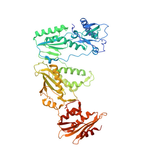
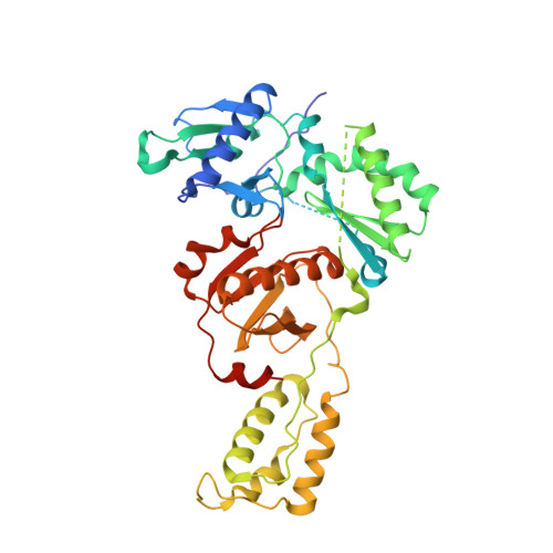
Elucidating molecular interactions ofL-nucleotides with HIV-1 reverse transcriptase and mechanism of M184V-caused drug resistance.

Hung, M.Tokarsky, E.J.Lagpacan, L.Zhang, L.Suo, Z.Lansdon, E.B.

(2019) Commun Biol 2: 469-469

  • DOI: https://doi.org/10.1038/s42003-019-0706-x
  • Primary Citation of Related Structures:  
    6UIR, 6UIS, 6UIT, 6UJX, 6UJY, 6UJZ, 6UK0

  • PubMed Abstract: 

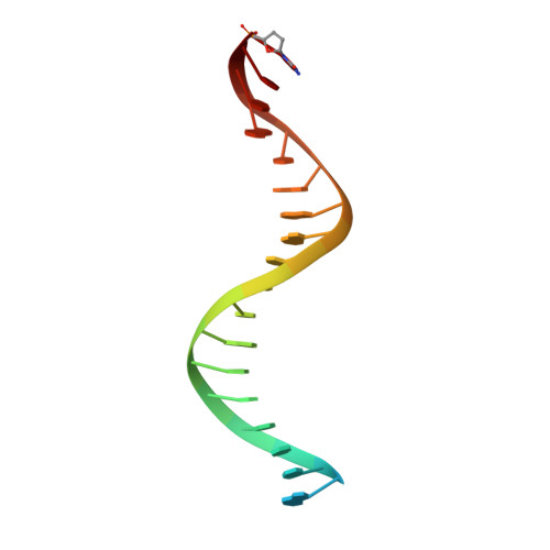
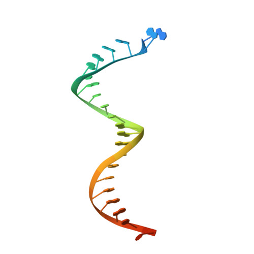
    Emtricitabine (FTC) and lamivudine (3TC), containing an oxathiolane ring with unnatural (-)-stereochemistry, are widely used nucleoside reverse transcriptase inhibitors (NRTIs) in anti-HIV therapy. Treatment with FTC or 3TC primarily selects for the HIV-1 RT M184V/I resistance mutations. Here we provide a comprehensive kinetic and structural basis for inhibiting HIV-1 RT by (-)-FTC-TP and (-)-3TC-TP and drug resistance by M184V. (-)-FTC-TP and (-)-3TC-TP have higher binding affinities (1/ K d ) for wild-type RT but slower incorporation rates than dCTP. HIV-1 RT ternary crystal structures with (-)-FTC-TP and (-)-3TC-TP corroborate kinetic results demonstrating that their oxathiolane sulfur orients toward the DNA primer 3'-terminus and their triphosphate exists in two different binding conformations. M184V RT displays greater (>200-fold) K d for the L -nucleotides and moderately higher (>9-fold) K d for the D -isomers compared to dCTP. The M184V RT structure illustrates how the mutation repositions the oxathiolane of (-)-FTC-TP and shifts its triphosphate into a non-productive conformation.


  • Organizational Affiliation

    1Gilead Sciences, Inc., 333 Lakeside Dr., Foster City, CA 94404 USA.


Macromolecules

Find similar proteins by:  (by identity cutoff)  |  3D Structure
Entity ID: 1
MoleculeChains Sequence LengthOrganismDetailsImage
p66 Reverse transcriptase/RNaseH572HIV-1 M:B_HXB2RMutation(s): 2 
Gene Names: gag-pol
EC: 3.4.23.16 (PDB Primary Data), 2.7.7.49 (PDB Primary Data), 2.7.7.7 (PDB Primary Data), 3.1.26.13 (PDB Primary Data), 3.1.13.2 (PDB Primary Data), 2.7.7 (PDB Primary Data), 3.1 (PDB Primary Data)
UniProt
Find proteins for P04585 (Human immunodeficiency virus type 1 group M subtype B (isolate HXB2))
Explore P04585 
Go to UniProtKB:  P04585
Entity Groups  
Sequence Clusters30% Identity50% Identity70% Identity90% Identity95% Identity100% Identity
UniProt GroupP04585
Sequence Annotations
Expand
  • Reference Sequence
Find similar proteins by:  (by identity cutoff)  |  3D Structure
Entity ID: 2
MoleculeChains Sequence LengthOrganismDetailsImage
p51 Reverse transcriptase/RNaseH440HIV-1 M:B_HXB2RMutation(s): 1 
Gene Names: gag-pol
EC: 3.4.23.16 (PDB Primary Data), 2.7.7.49 (PDB Primary Data), 2.7.7.7 (PDB Primary Data), 3.1.26.13 (PDB Primary Data), 3.1.13.2 (PDB Primary Data), 2.7.7 (PDB Primary Data), 3.1 (PDB Primary Data)
UniProt
Find proteins for P04585 (Human immunodeficiency virus type 1 group M subtype B (isolate HXB2))
Explore P04585 
Go to UniProtKB:  P04585
Entity Groups  
Sequence Clusters30% Identity50% Identity70% Identity90% Identity95% Identity100% Identity
UniProt GroupP04585
Sequence Annotations
Expand
  • Reference Sequence

Find similar nucleic acids by:  Sequence   |   3D Structure  

Entity ID: 3
MoleculeChains LengthOrganismImage
DNA primerC [auth P]21Human immunodeficiency virus 1
Sequence Annotations
Expand
  • Reference Sequence
Find similar nucleic acids by:  (by identity cutoff)  |  3D Structure
Entity ID: 4
MoleculeChains LengthOrganismImage
DNA templateD [auth T]27Human immunodeficiency virus 1
Sequence Annotations
Expand
  • Reference Sequence
Small Molecules
Binding Affinity Annotations 
IDSourceBinding Affinity
1RY BindingDB:  6UJX Kd: 1.20e+4 (nM) from 1 assay(s)
Experimental Data & Validation

Experimental Data

  • Method: X-RAY DIFFRACTION
  • Resolution: 2.70 Å
  • R-Value Free: 0.244 
  • R-Value Work: 0.197 
  • R-Value Observed: 0.199 
  • Space Group: C 2 2 21
Unit Cell:
Length ( Å )Angle ( ˚ )
a = 169.804α = 90
b = 167.685β = 90
c = 103.309γ = 90
Software Package:
Software NamePurpose
PHENIXrefinement
PHENIXrefinement
HKL-2000data reduction
HKL-2000data scaling
PHENIXphasing

Structure Validation

View Full Validation Report


Ligand Structure Quality Assessment 


Entry History 

Deposition Data

Revision History  (Full details and data files)

  • Version 1.0: 2019-12-25
    Type: Initial release
  • Version 1.1: 2020-01-08
    Changes: Database references
  • Version 1.2: 2023-10-11
    Changes: Data collection, Database references, Derived calculations, Refinement description