6UJP | pdb_00006ujp

P-glycoprotein mutant-F979A and C952A-with BDE100


Experimental Data Snapshot

  • Method: X-RAY DIFFRACTION
  • Resolution: 3.98 Å
  • R-Value Free: 
    0.263 (Depositor), 0.257 (DCC) 
  • R-Value Work: 
    0.239 (Depositor), 0.239 (DCC) 
  • R-Value Observed: 
    0.241 (Depositor) 

Starting Model: experimental
View more details

wwPDB Validation 3D Report Full Report

Validation slider image for 6UJP

Ligand Structure Quality Assessment 


This is version 1.2 of the entry. See complete history

Literature

Structural definition of polyspecific compensatory ligand recognition by P-glycoprotein.

Le, C.A.Harvey, D.S.Aller, S.G.

(2020) IUCrJ 7: 663-672

  • DOI: https://doi.org/10.1107/S2052252520005709
  • Primary Citation Related Structures: 
    6UJN, 6UJP, 6UJR, 6UJS, 6UJT, 6UJW

  • PubMed Abstract: 

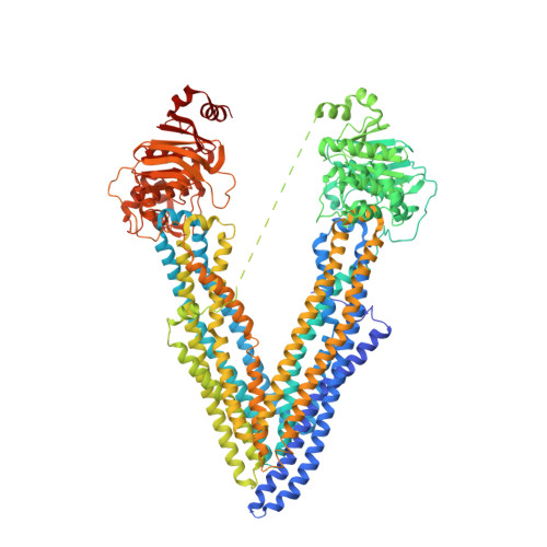
    The multidrug transporter P-glycoprotein (Pgp)/ABCB1/MDR1 plays an important role in multidrug resistance (MDR) and detoxification owing to its ability to efflux an unusually large and chemically diverse set of substrates. Previous phenylalanine-to-alanine scanning mutagenesis of Pgp revealed that nearly all mutations retained full MDR function and still permitted substrate transport. This suggests that either the loss of any single aromatic side chain did not affect the ligand-binding modes or that highly adaptive and compensatory drug recognition is an intrinsic property including ligand-binding shifts that preserve function. To explore this hypothesis, the ATPase function and crystallographic localization of five single-site mutations in which the native aromatic residue directly interacted with the environmental pollutant BDE-100, as shown in previous crystal structures, were tested. Two mutants, Y303A and Y306A, showed strong BDE-100 occupancy at the original site (site 1), but also revealed a novel site 2 located on the opposing pseudo-symmetric half of the drug-binding pocket (DBP). Surprisingly, the F724A mutant structure had no detectable binding in site 1 but exhibited a novel site shifted 11 Å from site 1. ATPase studies revealed shifts in ATPase kinetics for the five mutants, but otherwise indicated a catalytically active transporter that was inhibited by BDE-100, similar to wild-type Pgp. These results emphasize a high degree of compensatory drug recognition in Pgp that is made possible by aromatic amino-acid side chains concentrated in the DBP. Compensatory recognition forms the underpinning of polyspecific drug transport, but also highlights the challenges associated with the design of therapeutics that evade efflux altogether.


  • Organizational Affiliation
    • Department of Pharmacology and Toxicology, University of Alabama at Birmingham, Birmingham, AL 35294, USA.

Macromolecule Content 

  • Total Structure Weight: 142.13 kDa 
  • Atom Count: 9,175 
  • Modeled Residue Count: 1,182 
  • Deposited Residue Count: 1,282 
  • Unique protein chains: 1

Macromolecules

Find similar proteins by:|  3D Structure
Entity ID: 1
MoleculeChains  Sequence LengthOrganismDetailsImage
ATP-dependent translocase ABCB11,282Mus musculusMutation(s): 5 
Gene Names: Abcb1aAbcb4Mdr1aMdr3Pgy-3Pgy3
EC: 7.6.2.2 (PDB Primary Data), 7.6.2.1 (PDB Primary Data)
Membrane Entity: Yes 
UniProt
Find proteins for P21447 (Mus musculus)
Explore P21447 
Go to UniProtKB:  P21447
Entity Groups
Sequence Clusters30% Identity50% Identity70% Identity90% Identity95% Identity100% Identity
UniProt GroupP21447
Sequence Annotations
Expand
Reference Sequence

Small Molecules

Ligands 1 Unique
IDChains Name / Formula / InChI Key2D Diagram3D Interactions
4C8
(Subject of Investigation/LOI)

Query on 4C8


Download:Ideal Coordinates CCD File
B [auth A]2,4-dibromophenyl 2,4,6-tribromophenyl ether
C12 H5 Br5 O
NSKIRYMHNFTRLR-UHFFFAOYSA-N

Experimental Data & Validation

Experimental Data

  • Method: X-RAY DIFFRACTION
  • Resolution: 3.98 Å
  • R-Value Free:  0.263 (Depositor), 0.257 (DCC) 
  • R-Value Work:  0.239 (Depositor), 0.239 (DCC) 
  • R-Value Observed: 0.241 (Depositor) 
Space Group: P 21 21 21
Unit Cell:
Length ( Å )Angle ( ˚ )
a = 89.046α = 90
b = 138.133β = 90
c = 188.246γ = 90
Software Package:
Software NamePurpose
PHENIXrefinement
HKL-2000data scaling
PDB_EXTRACTdata extraction
HKL-2000data reduction
PHENIXphasing

Structure Validation

View Full Validation Report


Ligand Structure Quality Assessment 


Entry History 

Deposition Data

Revision History  (Full details and data files)

  • Version 1.0: 2020-05-27
    Type: Initial release
  • Version 1.1: 2020-08-05
    Changes: Database references
  • Version 1.2: 2023-10-11
    Changes: Data collection, Database references, Refinement description