


Open Dimer of Y77A Mutant Putative Ryanodine Receptor from Bacteroides thetaiotaomicron VPI-5482
Wu, R., Jedrzejczak, R., Joachimiak, A.To be published.
Experimental Data Snapshot
Entity ID: 1 | |||||
|---|---|---|---|---|---|
| Molecule | Chains | Sequence Length | Organism | Details | Image |
| Putative ryanodine receptor | 100 | Bacteroides thetaiotaomicron VPI-5482 | Mutation(s): 1 Gene Names: BT_2247 | 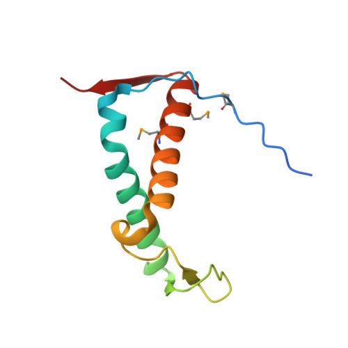 | |
UniProt | |||||
Entity Groups | |||||
| Sequence Clusters | 30% Identity50% Identity70% Identity90% Identity95% Identity100% Identity | ||||
| UniProt Group | Q8A5J2 | ||||
Sequence AnnotationsExpand | |||||
Reference Sequence | |||||
| Ligands 4 Unique | |||||
|---|---|---|---|---|---|
| ID | Chains | Name / Formula / InChI Key | 2D Diagram | 3D Interactions | |
| CFF (Subject of Investigation/LOI) Download:Ideal Coordinates CCD File | AA [auth F] BA [auth F] G [auth A] H [auth A] I [auth A] | CAFFEINE C8 H10 N4 O2 RYYVLZVUVIJVGH-UHFFFAOYSA-N |  | ||
| SO4 Download:Ideal Coordinates CCD File | K [auth A], L [auth A], N [auth B], T [auth D] | SULFATE ION O4 S QAOWNCQODCNURD-UHFFFAOYSA-L |  | ||
| GOL Download:Ideal Coordinates CCD File | CA [auth F] DA [auth F] EA [auth F] J [auth A] M [auth B] | GLYCEROL C3 H8 O3 PEDCQBHIVMGVHV-UHFFFAOYSA-N |  | ||
| PYR Download:Ideal Coordinates CCD File | U [auth D], V [auth D] | PYRUVIC ACID C3 H4 O3 LCTONWCANYUPML-UHFFFAOYSA-N |  | ||
| Modified Residues 1 Unique | |||||
|---|---|---|---|---|---|
| ID | Chains | Type | Formula | 2D Diagram | Parent |
| MSE Query on MSE | A, B, C, D, E A, B, C, D, E, F | L-PEPTIDE LINKING | C5 H11 N O2 Se |  | MET |
| Length ( Å ) | Angle ( ˚ ) |
|---|---|
| a = 84.201 | α = 90 |
| b = 84.201 | β = 90 |
| c = 229.704 | γ = 90 |
| Software Name | Purpose |
|---|---|
| PHENIX | refinement |
| SCALEPACK | data scaling |
| PDB_EXTRACT | data extraction |
| HKL-3000 | data reduction |
| HKL-3000 | phasing |


