


Crystal structure of the CBM3 from Bacillus subtilis at 1.28 angstrom resolution
Morais, M.A.B., Paiva, J.H., Murakami, M.T.To be published.
Experimental Data Snapshot
wwPDB Validation 3D Report Full Report

Entity ID: 1 | |||||
|---|---|---|---|---|---|
| Molecule | Chains | Sequence Length | Organism | Details | Image |
| Endoglucanase | 149 | Bacillus subtilis subsp. subtilis str. 168 | Mutation(s): 0 Gene Names: eglS, bglC, gld, BSU18130 EC: 3.2.1.4 | 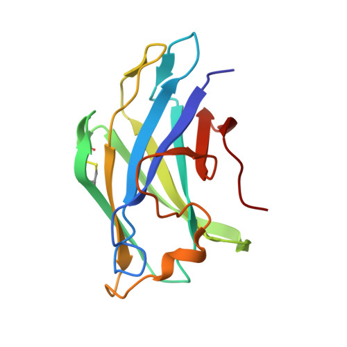 | |
UniProt | |||||
Entity Groups | |||||
| Sequence Clusters | 30% Identity50% Identity70% Identity90% Identity95% Identity100% Identity | ||||
| UniProt Group | P10475 | ||||
Sequence AnnotationsExpand | |||||
Reference Sequence | |||||
| Length ( Å ) | Angle ( ˚ ) |
|---|---|
| a = 51.092 | α = 90 |
| b = 55.176 | β = 90 |
| c = 89.427 | γ = 90 |
| Software Name | Purpose |
|---|---|
| PHENIX | refinement |
| XDS | data reduction |
| XSCALE | data scaling |
| PDB_EXTRACT | data extraction |
| Funding Organization | Location | Grant Number |
|---|---|---|
| Sao Paulo Research Foundation (FAPESP) | Brazil | 08/10555-1 |


