6UFE

The structure of a potassium selective ion channel at atomic resolution


Experimental Data Snapshot

  • Method: X-RAY DIFFRACTION
  • Resolution: 1.20 Å
  • R-Value Free: 0.148 
  • R-Value Work: 0.133 
  • R-Value Observed: 0.133 

wwPDB Validation   3D Report Full Report

Currently 6UFE does not have a validation slider image.


This is version 1.2 of the entry. See complete history


Literature

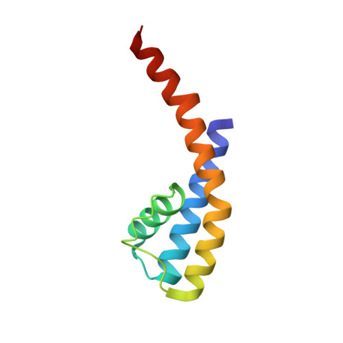
The structure of a potassium-selective ion channel reveals a hydrophobic gate regulating ion permeation.

Langan, P.S.Vandavasi, V.G.Kopec, W.Sullivan, B.Afonne, P.V.Weiss, K.L.de Groot, B.L.Coates, L.

(2020) IUCrJ 7: 835-843

  • DOI: https://doi.org/10.1107/S2052252520008271
  • Primary Citation of Related Structures:  
    6UFE

  • PubMed Abstract: 

    Protein dynamics are essential to function. One example of this is the various gating mechanisms within ion channels, which are transmembrane proteins that act as gateways into the cell. Typical ion channels switch between an open and closed state via a conformational transition which is often triggered by an external stimulus, such as ligand binding or pH and voltage differences. The atomic resolution structure of a potassium-selective ion channel named NaK2K has allowed us to observe that a hydro-phobic residue at the bottom of the selectivity filter, Phe92, appears in dual conformations. One of the two conformations of Phe92 restricts the diameter of the exit pore around the selectivity filter, limiting ion flow through the channel, while the other conformation of Phe92 provides a larger-diameter exit pore from the selectivity filter. Thus, it can be concluded that Phe92 acts as a hydro-phobic gate, regulating the flow of ions through the selectivity filter.


  • Organizational Affiliation

    Neutron Scattering Division, Oak Ridge National Laboratory, 1 Bethel Valley Road, Oak Ridge, TN 37831, USA.


Macromolecules
Find similar proteins by:  (by identity cutoff)  |  3D Structure
Entity ID: 1
MoleculeChains Sequence LengthOrganismDetailsImage
Transporter
A, B
92Bacillus cereusMutation(s): 2 
Gene Names: A9485_19160BACERE00184_02078CJ306_03585CN950_06075CN980_22870COI98_17615CON37_12595
Membrane Entity: Yes 
UniProt
Find proteins for Q81HW2 (Bacillus cereus (strain ATCC 14579 / DSM 31 / CCUG 7414 / JCM 2152 / NBRC 15305 / NCIMB 9373 / NCTC 2599 / NRRL B-3711))
Explore Q81HW2 
Go to UniProtKB:  Q81HW2
Entity Groups  
Sequence Clusters30% Identity50% Identity70% Identity90% Identity95% Identity100% Identity
UniProt GroupQ81HW2
Sequence Annotations
Expand
  • Reference Sequence
Small Molecules
Ligands 2 Unique
IDChains Name / Formula / InChI Key2D Diagram3D Interactions
MPD
Query on MPD

Download Ideal Coordinates CCD File 
C [auth A]
D [auth A]
E [auth A]
F [auth A]
K [auth B]
C [auth A],
D [auth A],
E [auth A],
F [auth A],
K [auth B],
L [auth B],
Q [auth B],
R [auth B],
S [auth B],
T [auth B]
(4S)-2-METHYL-2,4-PENTANEDIOL
C6 H14 O2
SVTBMSDMJJWYQN-YFKPBYRVSA-N
K
Query on K

Download Ideal Coordinates CCD File 
G [auth A]
H [auth A]
I [auth A]
J [auth A]
M [auth B]
G [auth A],
H [auth A],
I [auth A],
J [auth A],
M [auth B],
N [auth B],
O [auth B],
P [auth B]
POTASSIUM ION
K
NPYPAHLBTDXSSS-UHFFFAOYSA-N
Experimental Data & Validation

Experimental Data

  • Method: X-RAY DIFFRACTION
  • Resolution: 1.20 Å
  • R-Value Free: 0.148 
  • R-Value Work: 0.133 
  • R-Value Observed: 0.133 
  • Space Group: I 4
Unit Cell:
Length ( Å )Angle ( ˚ )
a = 67.86α = 90
b = 67.86β = 90
c = 89.62γ = 90
Software Package:
Software NamePurpose
PHENIXrefinement
XDSdata reduction
XSCALEdata scaling

Structure Validation

View Full Validation Report

Currently 6UFE does not have a validation slider image.


Entry History 

Deposition Data

Revision History  (Full details and data files)

  • Version 1.0: 2020-08-05
    Type: Initial release
  • Version 1.1: 2020-09-30
    Changes: Database references
  • Version 1.2: 2023-10-11
    Changes: Data collection, Database references, Refinement description