Structural Origins of Altered Spectroscopic Properties upon Ligand Binding in Proteins Containing a Fluorescent Noncanonical Amino Acid.
Gleason, P.R., Kolbaba-Kartchner, B., Henderson, J.N., Stahl, E.P., Simmons, C.R., Mills, J.H.(2021) Biochemistry 60: 2577-2585
- PubMed: 34415744 Search on PubMedSearch on PubMed Central
- DOI: https://doi.org/10.1021/acs.biochem.1c00291
- Primary Citation Related Structures:
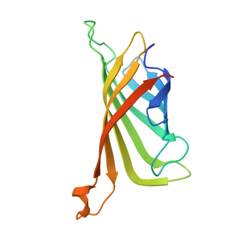
6UC3, 6UD1, 6UD6, 6UDB, 6UDC - PubMed Abstract:
Fluorescent noncanonical amino acids (fNCAAs) could serve as starting points for the rational design of protein-based fluorescent sensors of biological activity. However, efforts toward this goal are likely hampered by a lack of atomic-level characterization of fNCAAs within proteins. Here, we describe the spectroscopic and structural characterization of five streptavidin mutants that contain the fNCAA l-(7-hydroxycoumarin-4-yl)ethylglycine (7-HCAA) at sites proximal to the binding site of its substrate, biotin. Many of the mutants exhibited altered fluorescence spectra in response to biotin binding, which included both increases and decreases in fluorescence intensity as well as red- or blue-shifted emission maxima. Structural data were also obtained for three of the five mutants. The crystal structures shed light on interactions between 7-HCAA and functional groups, contributed either by the protein or by the substrate, that may be responsible for the observed changes in the 7-HCAA spectra. These data could be used in future studies aimed at the rational design of fluorescent, protein-based sensors of small molecule binding or dissociation.