6U7O | pdb_00006u7o

HIV-1 wild type protease with GRL-00819A, with phenyl-boronic-acid as P2'-ligand and with a 6-5-5-ring fused crown-like tetrahydropyranofuran as the P2-ligand


Experimental Data Snapshot

  • Method: X-RAY DIFFRACTION
  • Resolution: 1.33 Å
  • R-Value Free: 
    0.177 (Depositor), 0.142 (DCC) 
  • R-Value Work: 
    0.137 (Depositor), 0.145 (DCC) 
  • R-Value Observed: 
    0.139 (Depositor) 

Starting Model: experimental
View more details

wwPDB Validation 3D Report Full Report

Validation slider image for 6U7O

Ligand Structure Quality Assessment 


This is version 1.3 of the entry. See complete history

Literature

Potent HIV-1 Protease Inhibitors Containing Carboxylic and Boronic Acids: Effect on Enzyme Inhibition and Antiviral Activity and Protein-Ligand X-ray Structural Studies.

Ghosh, A.K.Xia, Z.Kovela, S.Robinson, W.L.Johnson, M.E.Kneller, D.W.Wang, Y.F.Aoki, M.Takamatsu, Y.Weber, I.T.Mitsuya, H.

(2019) ChemMedChem 14: 1863-1872

  • DOI: https://doi.org/10.1002/cmdc.201900508
  • Primary Citation Related Structures: 
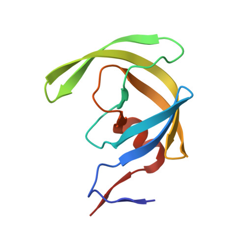
    6U7O, 6U7P

  • PubMed Abstract: 

    We report the synthesis and biological evaluation of phenylcarboxylic acid and phenylboronic acid containing HIV-1 protease inhibitors and their functional effect on enzyme inhibition and antiviral activity in MT-2 cell lines. Inhibitors bearing bis-THF ligand as P2 ligand and phenylcarboxylic acids and carboxamide as the P2' ligands, showed very potent HIV-1 protease inhibitory activity. However, carboxylic acid containing inhibitors showed very poor antiviral activity relative to carboxamide-derived inhibitors which showed good antiviral IC 50 value. Boronic acid derived inhibitor with bis-THF as the P2 ligand showed very potent enzyme inhibitory activity, but it showed lower antiviral activity than darunavir in the same assay. Boronic acid containing inhibitor with a P2-Crn-THF ligand also showed potent enzyme K i but significantly decreased antiviral activity. We have evaluated antiviral activity against a panel of highly drug-resistant HIV-1 variants. One of the inhibitors maintained good antiviral activity against HIV DRV R P20 and HIV DRV R P30 viruses. We have determined high resolution X-ray structures of two synthetic inhibitors bound to HIV-1 protease and obtained molecular insight into the ligand-binding site interactions.


  • Organizational Affiliation
    • Department of Chemistry and Department of Medicinal Chemistry, Purdue University, West Lafayette, IN, 47907, USA.

Macromolecule Content 

  • Total Structure Weight: 22.39 kDa 
  • Atom Count: 1,944 
  • Modeled Residue Count: 198 
  • Deposited Residue Count: 198 
  • Unique protein chains: 1

Macromolecules

Find similar proteins by:|  3D Structure
Entity ID: 1
MoleculeChains  Sequence LengthOrganismDetailsImage
Protease
A, B
99Human immunodeficiency virus 1Mutation(s): 5 
Gene Names: pol
UniProt
Find proteins for Q5RZ08 (Human immunodeficiency virus type 1)
Explore Q5RZ08 
Go to UniProtKB:  Q5RZ08
Entity Groups
Sequence Clusters30% Identity50% Identity70% Identity90% Identity95% Identity100% Identity
UniProt GroupQ5RZ08
Sequence Annotations
Expand
Reference Sequence

Small Molecules

Ligands 5 Unique
IDChains Name / Formula / InChI Key2D Diagram3D Interactions
Q1D
(Subject of Investigation/LOI)

Query on Q1D


Download:Ideal Coordinates CCD File
H [auth B]{4-[{(2R,3S)-3-[({[(3S,3aR,5R,7aS,8S)-hexahydro-4H-3,5-methanofuro[2,3-b]pyran-8-yl]oxy}carbonyl)amino]-2-hydroxy-4-phenylbutyl}(2-methylpropyl)sulfamoyl]phenyl}boronic acid
C29 H39 B N2 O9 S
QIKRDCAMSNJRAO-HGKZDDSVSA-N
GOL

Query on GOL


Download:Ideal Coordinates CCD File
G [auth A]GLYCEROL
C3 H8 O3
PEDCQBHIVMGVHV-UHFFFAOYSA-N
FMT

Query on FMT


Download:Ideal Coordinates CCD File
F [auth A]FORMIC ACID
C H2 O2
BDAGIHXWWSANSR-UHFFFAOYSA-N
CL

Query on CL


Download:Ideal Coordinates CCD File
D [auth A],
E [auth A],
I [auth B],
J [auth B]
CHLORIDE ION
Cl
VEXZGXHMUGYJMC-UHFFFAOYSA-M
NA

Query on NA


Download:Ideal Coordinates CCD File
C [auth A]SODIUM ION
Na
FKNQFGJONOIPTF-UHFFFAOYSA-N

Experimental Data & Validation

Experimental Data

  • Method: X-RAY DIFFRACTION
  • Resolution: 1.33 Å
  • R-Value Free:  0.177 (Depositor), 0.142 (DCC) 
  • R-Value Work:  0.137 (Depositor), 0.145 (DCC) 
  • R-Value Observed: 0.139 (Depositor) 
Space Group: P 21 21 2
Unit Cell:
Length ( Å )Angle ( ˚ )
a = 58.921α = 90
b = 86.479β = 90
c = 45.875γ = 90
Software Package:
Software NamePurpose
HKL-2000data scaling
PHASERphasing
REFMACrefinement
PDB_EXTRACTdata extraction
HKL-2000data reduction

Structure Validation

View Full Validation Report


Ligand Structure Quality Assessment 


Entry History 

& Funding Information

Deposition Data


Funding OrganizationLocationGrant Number
National Institutes of Health/National Human Genome Research Institute (NIH/NHGRI)United StatesAI150466
National Institutes of Health/National Cancer Institute (NIH/NCI)United StatesAI150461

Revision History  (Full details and data files)

  • Version 1.0: 2019-10-09
    Type: Initial release
  • Version 1.1: 2019-11-20
    Changes: Database references
  • Version 1.2: 2019-12-04
    Changes: Author supporting evidence
  • Version 1.3: 2023-10-11
    Changes: Data collection, Database references, Derived calculations, Refinement description