


To be published
Soule, J., Gnann, A.D., Parker, M.J., McKenna, K.C., Nguyen, S.V., Phan, N.T., Wicht, D.K., Dowling, D.P.To be published.
Experimental Data Snapshot
Starting Model: experimental
View more details
wwPDB Validation 3D Report Full Report

Entity ID: 1 | |||||
|---|---|---|---|---|---|
| Molecule | Chains | Sequence Length | Organism | Details | Image |
| methanesulfinate monooxygenase | 415 | Pseudomonas fluorescens Pf0-1 | Mutation(s): 0 Gene Names: Pfl01_3917 | 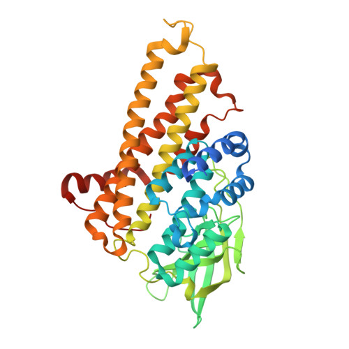 | |
UniProt | |||||
Entity Groups | |||||
| Sequence Clusters | 30% Identity50% Identity70% Identity90% Identity95% Identity100% Identity | ||||
| UniProt Group | Q3K9A0 | ||||
Sequence AnnotationsExpand | |||||
Reference Sequence | |||||
| Length ( Å ) | Angle ( ˚ ) |
|---|---|
| a = 80.554 | α = 90 |
| b = 162.411 | β = 90 |
| c = 61.285 | γ = 90 |
| Software Name | Purpose |
|---|---|
| PHENIX | refinement |
| HKL-2000 | data reduction |
| HKL-2000 | data scaling |
| PHASER | phasing |
| Coot | model building |
| Funding Organization | Location | Grant Number |
|---|---|---|
| National Science Foundation (NSF, United States) | United States | 1807480 |


