6U75 | pdb_00006u75

Crystal Structure of S. Cerevisiae SUMO E3 Ligase SIZ2


Experimental Data Snapshot

  • Method: X-RAY DIFFRACTION
  • Resolution: 2.63 Å
  • R-Value Free: 
    0.263 (Depositor), 0.256 (DCC) 
  • R-Value Work: 
    0.218 (Depositor) 
  • R-Value Observed: 
    0.220 (Depositor) 

Starting Model: experimental
View more details

wwPDB Validation 3D Report Full Report

Validation slider image for 6U75

This is version 1.2 of the entry. See complete history

Literature

DNA asymmetry promotes SUMO modification of the single-stranded DNA-binding protein RPA.

Cappadocia, L.Kochanczyk, T.Lima, C.D.

(2021) EMBO J 40: e103787-e103787

  • DOI: https://doi.org/10.15252/embj.2019103787
  • Primary Citation Related Structures: 
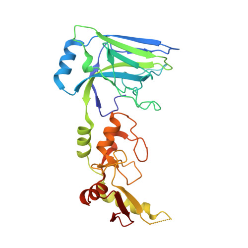
    6U75

  • PubMed Abstract: 

    Repair of DNA double-stranded breaks by homologous recombination (HR) is dependent on DNA end resection and on post-translational modification of repair factors. In budding yeast, single-stranded DNA is coated by replication protein A (RPA) following DNA end resection, and DNA-RPA complexes are then SUMO-modified by the E3 ligase Siz2 to promote repair. Here, we show using enzymatic assays that DNA duplexes containing 3' single-stranded DNA overhangs increase the rate of RPA SUMO modification by Siz2. The SAP domain of Siz2 binds DNA duplexes and makes a key contribution to this process as highlighted by models and a crystal structure of Siz2 and by assays performed using protein mutants. Enzymatic assays performed using DNA that can accommodate multiple RPA proteins suggest a model in which the SUMO-RPA signal is amplified by successive rounds of Siz2-dependent SUMO modification of RPA and dissociation of SUMO-RPA at the junction between single- and double-stranded DNA. Our results provide insights on how DNA architecture scaffolds a substrate and E3 ligase to promote SUMO modification in the context of DNA repair.


  • Organizational Affiliation
    • Structural Biology Program, Sloan Kettering Institute, New York, NY, USA.

Macromolecule Content 

  • Total Structure Weight: 61.65 kDa 
  • Atom Count: 4,237 
  • Modeled Residue Count: 522 
  • Deposited Residue Count: 538 
  • Unique protein chains: 1

Macromolecules

Find similar proteins by:|  3D Structure
Entity ID: 1
MoleculeChains  Sequence LengthOrganismDetailsImage
E3 SUMO-protein ligase SIZ2
A, B
269Saccharomyces cerevisiae S288CMutation(s): 0 
Gene Names: NFI1SIZ2YOR156C
EC: 2.3.2
UniProt
Find proteins for Q12216 (Saccharomyces cerevisiae (strain ATCC 204508 / S288c))
Explore Q12216 
Go to UniProtKB:  Q12216
Entity Groups
Sequence Clusters30% Identity50% Identity70% Identity90% Identity95% Identity100% Identity
UniProt GroupQ12216
Sequence Annotations
Expand
Reference Sequence

Experimental Data & Validation

Experimental Data

  • Method: X-RAY DIFFRACTION
  • Resolution: 2.63 Å
  • R-Value Free:  0.263 (Depositor), 0.256 (DCC) 
  • R-Value Work:  0.218 (Depositor) 
  • R-Value Observed: 0.220 (Depositor) 
Space Group: P 1 21 1
Unit Cell:
Length ( Å )Angle ( ˚ )
a = 61.916α = 90
b = 80.689β = 94.995
c = 76.527γ = 90
Software Package:
Software NamePurpose
PHENIXrefinement
HKL-2000data reduction
SCALEPACKdata scaling
PHENIXphasing

Structure Validation

View Full Validation Report


Entry History 

& Funding Information

Deposition Data


Funding OrganizationLocationGrant Number
National Institutes of Health/National Institute of General Medical Sciences (NIH/NIGMS)United StatesGM065872
National Institutes of Health/National Institute of General Medical Sciences (NIH/NIGMS)United StatesGM118080

Revision History  (Full details and data files)

  • Version 1.0: 2020-10-28
    Type: Initial release
  • Version 1.1: 2022-02-16
    Changes: Database references
  • Version 1.2: 2023-10-11
    Changes: Data collection, Refinement description