6U6U | pdb_00006u6u

IL36R extracellular domain in complex with BI655130 Fab


Experimental Data Snapshot

  • Method: X-RAY DIFFRACTION
  • Resolution: 2.31 Å
  • R-Value Free: 
    0.202 (Depositor), 0.205 (DCC) 
  • R-Value Work: 
    0.179 (Depositor), 0.185 (DCC) 
  • R-Value Observed: 
    0.180 (Depositor) 

Starting Model: experimental
View more details

wwPDB Validation 3D Report Full Report

Validation slider image for 6U6U

Ligand Structure Quality Assessment 


This is version 2.2 of the entry. See complete history

Literature

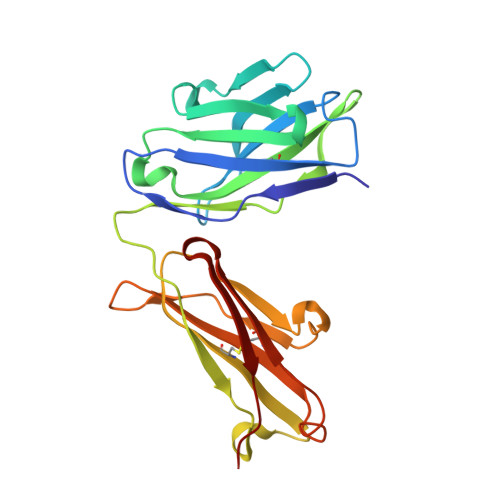
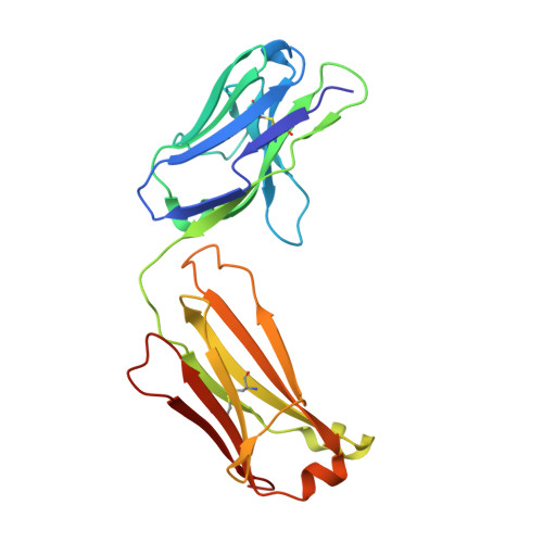
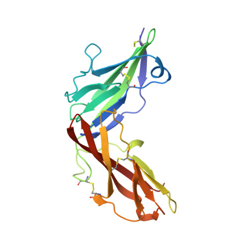
X-ray crystal structure localizes the mechanism of inhibition of an IL-36R antagonist monoclonal antibody to interaction with Ig1 and Ig2 extra cellular domains.

Larson, E.T.Brennan, D.L.Hickey, E.R.Ganesan, R.Kroe-Barrett, R.Farrow, N.A.

(2020) Protein Sci 29: 1679-1686

  • DOI: https://doi.org/10.1002/pro.3862
  • Primary Citation Related Structures: 
    6U6U

  • PubMed Abstract: 

    Cellular signaling via binding of the cytokines IL-36α, β, and γ along with binding of the accessory protein IL-36RAcP, to their cognate receptor IL-36R is believed to play a major role in epithelial and immune cell-mediated inflammation responses. Antagonizing the signaling cascade that results from these binding events via a directed monoclonal antibody provides an opportunity to suppress such immune responses. We report here the molecular structure of a complex between an extracellular portion of human IL-36R and a Fab derived from a high affinity anti-IL-36R neutralizing monoclonal antibody at 2.3 Å resolution. This structure, the first of IL-36R, reveals similarities with other structurally characterized IL-1R family members and elucidates the molecular determinants leading to the high affinity binding of the monoclonal antibody. The structure of the complex reveals that the epitope recognized by the Fab is remote from both the putative ligand and accessory protein binding interfaces on IL-36R, suggesting that the functional activity of the antibody is noncompetitive for these binding events.


  • Organizational Affiliation
    • Rheos Medicines, Cambridge, Massachusetts, USA.

Macromolecule Content 

  • Total Structure Weight: 70.48 kDa 
  • Atom Count: 5,225 
  • Modeled Residue Count: 632 
  • Deposited Residue Count: 633 
  • Unique protein chains: 3

Macromolecules

Find similar proteins by:|  3D Structure
Entity ID: 1
MoleculeChains  Sequence LengthOrganismDetailsImage
BI00655130 Fab heavy chainA [auth H]221Homo sapiensMutation(s): 0 
Find similar proteins by:|  3D Structure
Entity ID: 2
MoleculeChains  Sequence LengthOrganismDetailsImage
BI00655130 Fab light chainB [auth L]215Homo sapiensMutation(s): 0 
Find similar proteins by:|  3D Structure
Entity ID: 3
MoleculeChains  Sequence LengthOrganismDetailsImage
Interleukin-1 receptor-like 2C [auth R]197Homo sapiensMutation(s): 0 
Gene Names: IL1RL2IL1RRP2
EC: 3.2.2.6
UniProt & NIH Common Fund Data Resources
Find proteins for Q9HB29 (Homo sapiens)
Explore Q9HB29 
Go to UniProtKB:  Q9HB29
PHAROS:  Q9HB29
GTEx:  ENSG00000115598 
Entity Groups
Sequence Clusters30% Identity50% Identity70% Identity90% Identity95% Identity100% Identity
UniProt GroupQ9HB29
Glycosylation
Glycosylation Sites: 2Go to GlyGen: Q9HB29-1
Sequence Annotations
Expand
Reference Sequence

Oligosaccharides

Help  
Entity ID: 4
MoleculeChains Length2D Diagram GlycosylationD Interactions
2-acetamido-2-deoxy-beta-D-glucopyranose-(1-4)-2-acetamido-2-deoxy-beta-D-glucopyranoseD [auth A]2N-Glycosylation
Glycosylation Resources
GlyTouCan: G42666HT
GlyCosmos: G42666HT
GlyGen: G42666HT

Small Molecules

Experimental Data & Validation

Experimental Data

  • Method: X-RAY DIFFRACTION
  • Resolution: 2.31 Å
  • R-Value Free:  0.202 (Depositor), 0.205 (DCC) 
  • R-Value Work:  0.179 (Depositor), 0.185 (DCC) 
  • R-Value Observed: 0.180 (Depositor) 
Space Group: P 21 21 21
Unit Cell:
Length ( Å )Angle ( ˚ )
a = 60.698α = 90
b = 70.663β = 90
c = 233.268γ = 90
Software Package:
Software NamePurpose
XDSdata reduction
d*TREKdata scaling
PHASERphasing
PHENIXrefinement
PDB_EXTRACTdata extraction

Structure Validation

View Full Validation Report


Ligand Structure Quality Assessment 


Entry History 

Deposition Data

Revision History  (Full details and data files)

  • Version 1.0: 2020-04-22
    Type: Initial release
  • Version 1.1: 2020-05-27
    Changes: Database references
  • Version 1.2: 2020-07-08
    Changes: Database references
  • Version 2.0: 2020-07-29
    Type: Remediation
    Reason: Carbohydrate remediation
    Changes: Atomic model, Data collection, Derived calculations, Structure summary
  • Version 2.1: 2023-10-11
    Changes: Data collection, Database references, Refinement description, Structure summary
  • Version 2.2: 2024-10-23
    Changes: Structure summary