Probing transient excited states of the bacterial cell division regulator MinE by relaxation dispersion NMR spectroscopy.
Cai, M., Huang, Y., Shen, Y., Li, M., Mizuuchi, M., Ghirlando, R., Mizuuchi, K., Clore, G.M.(2019) Proc Natl Acad Sci U S A 116: 25446-25455
- PubMed: 31772021 Search on PubMedSearch on PubMed Central
- DOI: https://doi.org/10.1073/pnas.1915948116
- Primary Citation Related Structures:
6U6P, 6U6Q, 6U6R, 6U6S - PubMed Abstract:
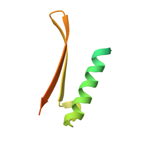
Bacterial MinD and MinE form a standing oscillatory wave which positions the cell division inhibitor MinC, that binds MinD, everywhere on the membrane except at the midpoint of the cell, ensuring midcell positioning of the cytokinetic septum. During this process MinE undergoes fold switching as it interacts with different partners. We explore the exchange dynamics between major and excited states of the MinE dimer in 3 forms using 15 N relaxation dispersion NMR: the full-length protein (6-stranded β-sheet sandwiched between 4 helices) representing the resting state; a 10-residue N-terminal deletion (Δ10) mimicking the membrane-binding competent state where the N-terminal helix is detached to interact with membrane; and N-terminal deletions of either 30 (Δ30) or 10 residues with an I24N mutation (Δ10/I24N), in which the β1-strands at the dimer interface are extruded and available to bind MinD, leaving behind a 4-stranded β-sheet. Full-length MinE samples 2 "excited" states: The first is similar to a full-length/Δ10 heterodimer; the second, also sampled by Δ10, is either similar to or well along the pathway toward the 4-stranded β-sheet form. Both Δ30 and Δ10/I24N sample 2 excited species: The first may involve destabilization of the β3- and β3'-strands at the dimer interface; changes in the second are more extensive, involving further disruption of secondary structure, possibly representing an ensemble of states on the pathway toward restoration of the resting state. The quantitative information on MinE conformational dynamics involving these excited states is crucial for understanding the oscillation pattern self-organization by MinD-MinE interaction dynamics on the membrane.
- Laboratory of Chemical Physics, National Institute of Diabetes and Digestive and Kidney Diseases, National Institutes of Health, Bethesda, MD 20892-0520.
Organizational Affiliation: