6U6B | pdb_00006u6b

Structure of human DNA polymerase beta misinserting dAMPNPP opposite the 5'G of the cisplatin Pt-GG intrastrand crosslink with Manganese in the active site


Experimental Data Snapshot

  • Method: X-RAY DIFFRACTION
  • Resolution: 3.11 Å
  • R-Value Free: 
    0.268 (Depositor), 0.265 (DCC) 
  • R-Value Work: 
    0.222 (Depositor), 0.220 (DCC) 
  • R-Value Observed: 
    0.224 (Depositor) 

Starting Model: experimental
View more details

wwPDB Validation 3D Report Full Report

Validation slider image for 6U6B

Ligand Structure Quality Assessment 


This is version 1.2 of the entry. See complete history

Literature

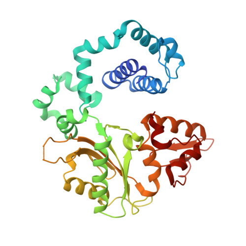
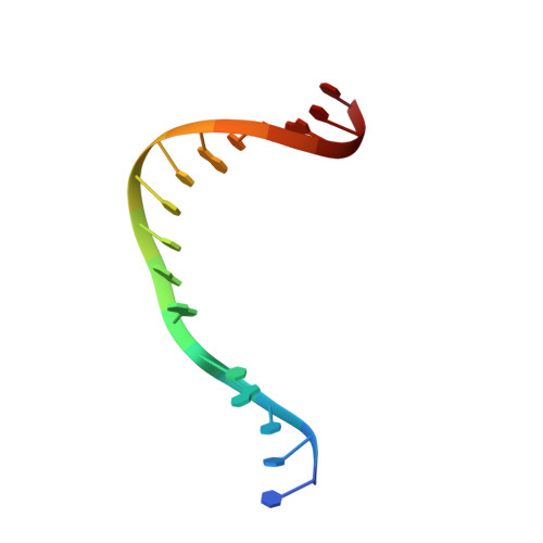
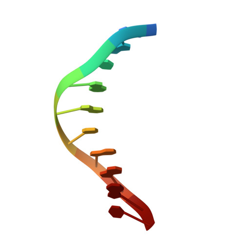
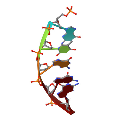
Structural insights into the promutagenic bypass of the major cisplatin-induced DNA lesion.

Ouzon-Shubeita, H.Vilas, C.K.Lee, S.

(2020) Biochem J 477: 937-951

  • DOI: https://doi.org/10.1042/BCJ20190906
  • Primary Citation Related Structures: 
    6U6B

  • PubMed Abstract: 

    The cisplatin-1,2-d(GpG) (Pt-GG) intrastrand cross-link is the predominant DNA lesion generated by cisplatin. Cisplatin has been shown to predominantly induce G to T mutations and Pt-GG permits significant misincorporation of dATP by human DNA polymerase β (polβ). In agreement, polβ overexpression, which is frequently observed in cancer cells, is linked to cisplatin resistance and a mutator phenotype. However, the structural basis for the misincorporation of dATP opposite Pt-GG is unknown. Here, we report the first structures of a DNA polymerase inaccurately bypassing Pt-GG. We solved two structures of polβ misincorporating dATP opposite the 5'-dG of Pt-GG in the presence of Mg2+ or Mn2+. The Mg2+-bound structure exhibits a sub-optimal conformation for catalysis, while the Mn2+-bound structure is in a catalytically more favorable semi-closed conformation. In both structures, dATP does not form a coplanar base pairing with Pt-GG. In the polβ active site, the syn-dATP opposite Pt-GG appears to be stabilized by protein templating and pi stacking interactions, which resembles the polβ-mediated dATP incorporation opposite an abasic site. Overall, our results suggest that the templating Pt-GG in the polβ active site behaves like an abasic site, promoting the insertion of dATP in a non-instructional manner.


  • Organizational Affiliation
    • Division of Chemical Biology and Medicinal Chemistry, College of Pharmacy, The University of Texas at Austin, Austin, TX 78712, U.S.A.

Macromolecule Content 

  • Total Structure Weight: 48.65 kDa 
  • Atom Count: 3,243 
  • Modeled Residue Count: 356 
  • Deposited Residue Count: 366 
  • Unique protein chains: 1
  • Unique nucleic acid chains: 3

Macromolecules


Find similar proteins by:|  3D Structure
Entity ID: 1
MoleculeChains  Sequence LengthOrganismDetailsImage
DNA polymerase beta335Homo sapiensMutation(s): 0 
Gene Names: POLB
EC: 2.7.7.7 (PDB Primary Data), 4.2.99 (PDB Primary Data), 4.2.99.18 (UniProt)
UniProt & NIH Common Fund Data Resources
Find proteins for P06746 (Homo sapiens)
Explore P06746 
Go to UniProtKB:  P06746
PHAROS:  P06746
GTEx:  ENSG00000070501 
Entity Groups
Sequence Clusters30% Identity50% Identity70% Identity90% Identity95% Identity100% Identity
UniProt GroupP06746
Sequence Annotations
Expand
Reference Sequence
Find similar nucleic acids by:  Sequence
Entity ID: 2
MoleculeChains LengthOrganismImage
DNA (5'-D(*CP*CP*CP*AP*CP*GP*GP*CP*CP*CP*AP*TP*CP*AP*CP*C)-3')B [auth T]16Homo sapiens
Sequence Annotations
Expand
Reference Sequence
Find similar nucleic acids by:  Sequence
Entity ID: 3
MoleculeChains LengthOrganismImage
DNA (5'-D(P*GP*GP*TP*GP*AP*TP*GP*GP*GP*C)-3')C [auth P]10Homo sapiens
Sequence Annotations
Expand
Reference Sequence
Find similar nucleic acids by:  Sequence
Entity ID: 4
MoleculeChains LengthOrganismImage
DNA (5'-D(P*GP*TP*GP*GP*G)-3')5Homo sapiens
Sequence Annotations
Expand
Reference Sequence

Small Molecules

Ligands 4 Unique
IDChains Name / Formula / InChI Key2D Diagram3D Interactions
DZ4

Query on DZ4


Download:Ideal Coordinates CCD File
I [auth A]2'-deoxy-5'-O-[(R)-hydroxy{[(R)-hydroxy(phosphonooxy)phosphoryl]amino}phosphoryl]adenosine
C10 H17 N6 O11 P3
WKIPJDSLGCBQCU-RRKCRQDMSA-N
CPT

Query on CPT


Download:Ideal Coordinates CCD File
J [auth T]Cisplatin
Cl2 H6 N2 Pt
LXZZYRPGZAFOLE-UHFFFAOYSA-L
MN

Query on MN


Download:Ideal Coordinates CCD File
E [auth A],
F [auth A]
MANGANESE (II) ION
Mn
WAEMQWOKJMHJLA-UHFFFAOYSA-N
NA

Query on NA


Download:Ideal Coordinates CCD File
G [auth A],
H [auth A]
SODIUM ION
Na
FKNQFGJONOIPTF-UHFFFAOYSA-N

Experimental Data & Validation

Experimental Data

  • Method: X-RAY DIFFRACTION
  • Resolution: 3.11 Å
  • R-Value Free:  0.268 (Depositor), 0.265 (DCC) 
  • R-Value Work:  0.222 (Depositor), 0.220 (DCC) 
  • R-Value Observed: 0.224 (Depositor) 
Space Group: P 1 21 1
Unit Cell:
Length ( Å )Angle ( ˚ )
a = 53.43α = 90
b = 78.216β = 110.57
c = 53.965γ = 90
Software Package:
Software NamePurpose
PHENIXrefinement
PDB_EXTRACTdata extraction
HKL-2000data scaling
HKL-2000data reduction
REFMACphasing

Structure Validation

View Full Validation Report


Ligand Structure Quality Assessment 


Entry History 

& Funding Information

Deposition Data


Funding OrganizationLocationGrant Number
National Institutes of Health/National Institute of Environmental Health Sciences (NIH/NIEHS)United StatesES26676

Revision History  (Full details and data files)

  • Version 1.0: 2020-09-09
    Type: Initial release
  • Version 1.1: 2021-03-24
    Changes: Database references
  • Version 1.2: 2023-10-11
    Changes: Data collection, Database references, Refinement description