6U4N | pdb_00006u4n

Solution structure of paxillin LIM4 in complex with kindlin-2 F0


Experimental Data Snapshot

  • Method: SOLUTION NMR
  • Conformers Calculated: 100 
  • Conformers Submitted: 20 
  • Selection Criteria: structures with the lowest energy 

wwPDB Validation 3D Report Full Report

Validation slider image for 6U4N

This is version 1.3 of the entry. See complete history

Literature

Structural Basis of Paxillin Recruitment by Kindlin-2 in Regulating Cell Adhesion.

Zhu, L.Liu, H.Lu, F.Yang, J.Byzova, T.V.Qin, J.

(2019) Structure 27: 1686

  • DOI: https://doi.org/10.1016/j.str.2019.09.006
  • Primary Citation Related Structures: 
    6U4M, 6U4N

  • PubMed Abstract: 

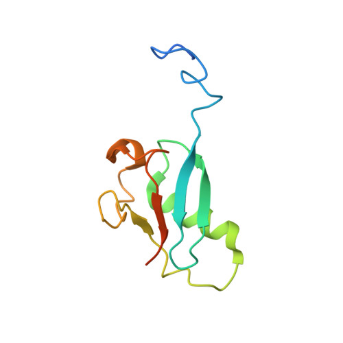
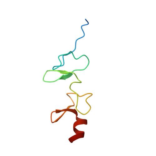
    Activation of cell surface receptor integrin has been extensively studied as the first key step to trigger cell adhesion, but the subsequent events, widely regarded as integrin "outside-in" signaling to form supramolecular complexes (focal adhesions [FAs]) to promote dynamic cell adhesion, remain poorly elucidated. Integrin activator kindlin-2 was recently found to associate with paxillin in nascent FAs, implicating an early yet undefined integrin outside-in signaling event. Here we show structurally that kindlin-2 recognizes paxillin via a distinct interface involving the ubiquitin-like kindlin-2 F0 domain and the paxillin LIM4 domain. The interface is adjacent to the membrane binding site of kindlin-2 F0, suggesting a mechanism for kindlin-2 to recruit paxillin to the membrane-proximal site where FA assembly is initiated. Disruption of the interface impaired the localization of paxillin, causing strong defects in FA assembly and cell migration. These data unveil a structural basis of the kindlin-2/paxillin interaction in controlling dynamic cell adhesion.


  • Organizational Affiliation
    • Department of Cardiovascular & Metabolic Sciences, Lerner Research Institute, Cleveland Clinic, 9500 Euclid Avenue, Cleveland, OH 44195, USA.

Macromolecule Content 

  • Total Structure Weight: 21.44 kDa 
  • Atom Count: 1,294 
  • Modeled Residue Count: 159 
  • Deposited Residue Count: 184 
  • Unique protein chains: 2

Macromolecules

Find similar proteins by:|  3D Structure
Entity ID: 1
MoleculeChains  Sequence LengthOrganismDetailsImage
Fermitin family homolog 2112Homo sapiensMutation(s): 0 
Gene Names: FERMT2KIND2MIG2PLEKHC1
UniProt & NIH Common Fund Data Resources
Find proteins for Q96AC1 (Homo sapiens)
Explore Q96AC1 
Go to UniProtKB:  Q96AC1
PHAROS:  Q96AC1
GTEx:  ENSG00000073712 
Entity Groups
Sequence Clusters30% Identity50% Identity70% Identity90% Identity95% Identity100% Identity
UniProt GroupQ96AC1
Sequence Annotations
Expand
Reference Sequence
Find similar proteins by:|  3D Structure
Entity ID: 2
MoleculeChains  Sequence LengthOrganismDetailsImage
Paxillin72Homo sapiensMutation(s): 0 
Gene Names: PXN
UniProt & NIH Common Fund Data Resources
Find proteins for P49023 (Homo sapiens)
Explore P49023 
Go to UniProtKB:  P49023
PHAROS:  P49023
GTEx:  ENSG00000089159 
Entity Groups
Sequence Clusters30% Identity50% Identity70% Identity90% Identity95% Identity100% Identity
UniProt GroupP49023
Sequence Annotations
Expand
Reference Sequence

Small Molecules

Experimental Data & Validation

Experimental Data

  • Method: SOLUTION NMR
  • Conformers Calculated: 100 
  • Conformers Submitted: 20 
  • Selection Criteria: structures with the lowest energy 

Structure Validation

View Full Validation Report


Entry History 

& Funding Information

Deposition Data


Funding OrganizationLocationGrant Number
National Institutes of Health/National Human Genome Research Institute (NIH/NHGRI)United States--

Revision History  (Full details and data files)

  • Version 1.0: 2019-10-23
    Type: Initial release
  • Version 1.1: 2019-11-20
    Changes: Database references
  • Version 1.2: 2019-12-18
    Changes: Author supporting evidence
  • Version 1.3: 2024-05-01
    Changes: Data collection, Database references