


Structure of Tn IYD bound in the semiquinone form bound to FMN and 3-iodo-L-tyrosine
Sun, Z., Kavran, J.M., Rokita, S.E.To be published.
Experimental Data Snapshot
Entity ID: 1 | |||||
|---|---|---|---|---|---|
| Molecule | Chains | Sequence Length | Organism | Details | Image |
| iodotyrosine deiodinase | 192 | Thermotoga neapolitana DSM 4359 | Mutation(s): 0 Gene Names: CTN_0569 EC: 1.21.1.1 | 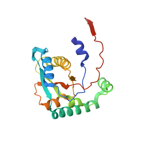 | |
UniProt | |||||
Entity Groups | |||||
| Sequence Clusters | 30% Identity50% Identity70% Identity90% Identity95% Identity100% Identity | ||||
| UniProt Group | B9K712 | ||||
Sequence AnnotationsExpand | |||||
Reference Sequence | |||||
| Ligands 3 Unique | |||||
|---|---|---|---|---|---|
| ID | Chains | Name / Formula / InChI Key | 2D Diagram | 3D Interactions | |
| FMN (Subject of Investigation/LOI) Download:Ideal Coordinates CCD File | C [auth A], J [auth B] | FLAVIN MONONUCLEOTIDE C17 H21 N4 O9 P FVTCRASFADXXNN-SCRDCRAPSA-N |  | ||
| IYR (Subject of Investigation/LOI) Download:Ideal Coordinates CCD File | D [auth A], K [auth B], N [auth B] | 3-IODO-TYROSINE C9 H10 I N O3 UQTZMGFTRHFAAM-ZETCQYMHSA-N |  | ||
| CL Download:Ideal Coordinates CCD File | E [auth A] F [auth A] G [auth A] H [auth A] I [auth A] | CHLORIDE ION Cl VEXZGXHMUGYJMC-UHFFFAOYSA-M |  | ||
| Length ( Å ) | Angle ( ˚ ) |
|---|---|
| a = 43.035 | α = 90 |
| b = 81.176 | β = 90 |
| c = 104.146 | γ = 90 |
| Software Name | Purpose |
|---|---|
| PHENIX | refinement |
| Aimless | data scaling |
| PDB_EXTRACT | data extraction |
| XDS | data reduction |
| PHENIX | phasing |
| Funding Organization | Location | Grant Number |
|---|---|---|
| National Science Foundation (NSF, United States) | United States | -- |


