Crystal structures of native cytochrome c6from Thermosynechococcus elongatus in two different space groups and implications for its oligomerization.
Falke, S., Feiler, C., Chapman, H., Sarrou, I.(2020) Acta Crystallogr F Struct Biol Commun 76: 444-452
- PubMed: 32880593 Search on PubMedSearch on PubMed Central
- DOI: https://doi.org/10.1107/S2053230X20010249
- Primary Citation Related Structures:
6TR1, 6TSY - PubMed Abstract:
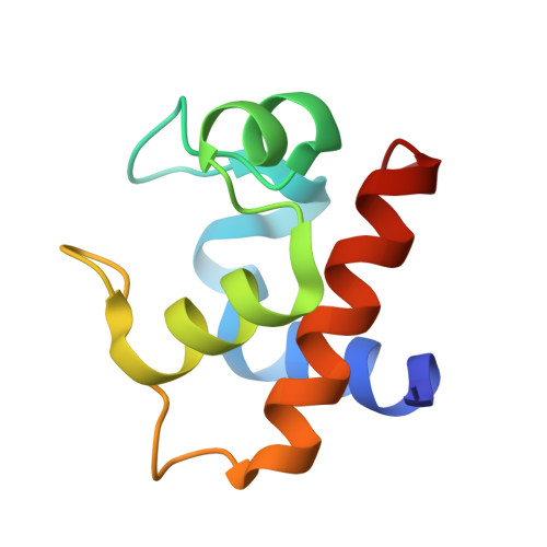
Native cytochrome c 6 was purified from an extract of strain BP-1 of the thermophilic cyanobacterium Thermosynechococcus elongatus. The protein was crystallized, and with only slight modifications of the buffer and vapour-diffusion conditions two different space groups were observed, namely H3 and C2. Both crystal structures were solved; they contained three and six molecules per asymmetric unit and were refined to 1.7 and 2.25 Å resolution, respectively. To date, the structure of native cytochrome c 6 from T. elongatus has only been reported as a monomer using NMR spectroscopy, i.e. without addressing putative oligomerization, and related structures have only previously been solved using X-ray crystallography after recombinant gene overexpression in Escherichia coli. The reported space groups of related cyanobacterial cytochrome c 6 structures differ from those reported here. Interestingly, the protein-protein interfaces that were observed utilizing X-ray crystallography could also explain homo-oligomerization in solution; specifically, trimerization is indicated by infra-red dynamic light scattering and blue native gel electrophoresis in solution. Trimers were also detected by mass spectrometry. Furthermore, there is an indication of post-translational methylation in the crystal structure. Additionally, the possibility of modifying the crystal size and the redox activity in the context of photosynthesis is shaping the investigated cytochrome as a highly suitable model protein for advanced serial crystallography at highly brilliant X-ray free-electron laser sources.
- Institute for Biochemistry and Molecular Biology, University of Hamburg, c/o DESY, Notkestrasse 85, 22603 Hamburg, Germany.
Organizational Affiliation: