6TR5

Melatonin-Notum complex

  • Classification: HYDROLASE
  • Organism(s): Homo sapiens
  • Expression System: Homo sapiens
  • Mutation(s): Yes 

  • Deposited: 2019-12-17 Released: 2020-01-15 
  • Deposition Author(s): Zhao, Y., Jones, E.Y.
  • Funding Organization(s): Medical Research Council (MRC, United Kingdom), Cancer Research UK

Experimental Data Snapshot

  • Method: X-RAY DIFFRACTION
  • Resolution: 1.51 Å
  • R-Value Free: 0.226 
  • R-Value Work: 0.196 
  • R-Value Observed: 0.198 

wwPDB Validation   3D Report Full Report


Ligand Structure Quality Assessment 


This is version 1.3 of the entry. See complete history


Literature

Structural characterization of melatonin as an inhibitor of the Wnt deacylase Notum.

Zhao, Y.Ren, J.Hillier, J.Jones, M.Lu, W.Jones, E.Y.

(2020) J Pineal Res 68: e12630-e12630

  • DOI: https://doi.org/10.1111/jpi.12630
  • Primary Citation of Related Structures:  
    6TR5, 6TR6, 6TR7

  • PubMed Abstract: 

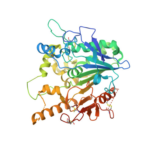
    The hormone melatonin, secreted from the pineal gland, mediates multiple physiological effects including modulation of Wnt/β-catenin signalling. The Wnt palmitoleate lipid modification is essential for its signalling activity, while the carboxylesterase Notum can remove the lipid from Wnt and inactivate it. Notum enzyme inhibition can therefore upregulate Wnt signalling. While searching for Notum inhibitors by crystallographic fragment screening, a hit compound N-[2-(5-fluoro-1H-indol-3-yl)ethyl]acetamide that is structurally similar to melatonin came to our attention. We then soaked melatonin and its precursor N-acetylserotonin into Notum crystals and obtained high-resolution structures (≤1.5 Å) of their complexes. In each of the structures, two compound molecules bind with Notum: one at the enzyme's catalytic pocket, overlapping the space occupied by the acyl tail of the Wnt palmitoleate lipid, and the other at the edge of the pocket opposite the substrate entrance. Although the inhibitory activity of melatonin shown by in vitro enzyme assays is low (IC 50 75 µmol/L), the structural information reported here provides a basis for the design of potent and brain accessible drugs for neurodegenerative diseases such as Alzheimer's disease, in which upregulation of Wnt signalling may be beneficial.


  • Organizational Affiliation

    Division of Structural Biology, Wellcome Centre for Human Genetics, University of Oxford, Oxford, UK.


Macromolecules
Find similar proteins by:  (by identity cutoff)  |  3D Structure
Entity ID: 1
MoleculeChains Sequence LengthOrganismDetailsImage
Palmitoleoyl-protein carboxylesterase NOTUM383Homo sapiensMutation(s): 1 
Gene Names: NOTUMOK/SW-CL.30
EC: 3.1.1.98
UniProt & NIH Common Fund Data Resources
Find proteins for Q6P988 (Homo sapiens)
Explore Q6P988 
Go to UniProtKB:  Q6P988
PHAROS:  Q6P988
GTEx:  ENSG00000185269 
Entity Groups  
Sequence Clusters30% Identity50% Identity70% Identity90% Identity95% Identity100% Identity
UniProt GroupQ6P988
Sequence Annotations
Expand
  • Reference Sequence
Small Molecules
Ligands 4 Unique
IDChains Name / Formula / InChI Key2D Diagram3D Interactions
ML1 (Subject of Investigation/LOI)
Query on ML1

Download Ideal Coordinates CCD File 
B [auth A],
C [auth A]
N-[2-(5-methoxy-1H-indol-3-yl)ethyl]acetamide
C13 H16 N2 O2
DRLFMBDRBRZALE-UHFFFAOYSA-N
NAG
Query on NAG

Download Ideal Coordinates CCD File 
E [auth A]2-acetamido-2-deoxy-beta-D-glucopyranose
C8 H15 N O6
OVRNDRQMDRJTHS-FMDGEEDCSA-N
SO4
Query on SO4

Download Ideal Coordinates CCD File 
F [auth A]
G [auth A]
H [auth A]
I [auth A]
J [auth A]
F [auth A],
G [auth A],
H [auth A],
I [auth A],
J [auth A],
K [auth A],
L [auth A],
M [auth A]
SULFATE ION
O4 S
QAOWNCQODCNURD-UHFFFAOYSA-L
DMS
Query on DMS

Download Ideal Coordinates CCD File 
D [auth A]DIMETHYL SULFOXIDE
C2 H6 O S
IAZDPXIOMUYVGZ-UHFFFAOYSA-N
Binding Affinity Annotations 
IDSourceBinding Affinity
ML1 Binding MOAD:  6TR5 IC50: 7.50e+4 (nM) from 1 assay(s)
Experimental Data & Validation

Experimental Data

  • Method: X-RAY DIFFRACTION
  • Resolution: 1.51 Å
  • R-Value Free: 0.226 
  • R-Value Work: 0.196 
  • R-Value Observed: 0.198 
  • Space Group: P 21 21 21
Unit Cell:
Length ( Å )Angle ( ˚ )
a = 60.259α = 90
b = 73.165β = 90
c = 79.001γ = 90
Software Package:
Software NamePurpose
PHENIXrefinement
PDB_EXTRACTdata extraction
xia2data reduction
xia2data scaling
xia2phasing

Structure Validation

View Full Validation Report


Ligand Structure Quality Assessment 


Entry History & Funding Information

Deposition Data


Funding OrganizationLocationGrant Number
Medical Research Council (MRC, United Kingdom)United KingdomMR/M000141/1
Cancer Research UKUnited KingdomC375/A17721

Revision History  (Full details and data files)

  • Version 1.0: 2020-01-15
    Type: Initial release
  • Version 1.1: 2020-02-19
    Changes: Data collection, Database references
  • Version 1.2: 2020-07-29
    Type: Remediation
    Reason: Carbohydrate remediation
    Changes: Data collection, Derived calculations, Structure summary
  • Version 1.3: 2024-01-24
    Changes: Data collection, Database references, Refinement description, Structure summary