6TMU

Crystal structure of the chaperonin gp146 from the bacteriophage EL 2 (Pseudomonas aeruginosa) in presence of ATP-BeFx, crystal form II


Experimental Data Snapshot

  • Method: X-RAY DIFFRACTION
  • Resolution: 3.54 Å
  • R-Value Free: 0.275 
  • R-Value Work: 0.250 
  • R-Value Observed: 0.252 

wwPDB Validation   3D Report Full Report


Ligand Structure Quality Assessment 


This is version 1.1 of the entry. See complete history


Literature

Structure and conformational cycle of a bacteriophage-encoded chaperonin.

Bracher, A.Paul, S.S.Wang, H.Wischnewski, N.Hartl, F.U.Hayer-Hartl, M.

(2020) PLoS One 15: e0230090-e0230090

  • DOI: https://doi.org/10.1371/journal.pone.0230090
  • Primary Citation of Related Structures:  
    6TMT, 6TMU, 6TMV, 6TMW, 6TMX

  • PubMed Abstract: 

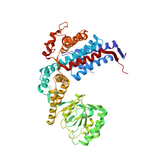
    Chaperonins are ubiquitous molecular chaperones found in all domains of life. They form ring-shaped complexes that assist in the folding of substrate proteins in an ATP-dependent reaction cycle. Key to the folding cycle is the transient encapsulation of substrate proteins by the chaperonin. Here we present a structural and functional characterization of the chaperonin gp146 (ɸEL) from the phage EL of Pseudomonas aeruginosa. ɸEL, an evolutionarily distant homolog of bacterial GroEL, is active in ATP hydrolysis and prevents the aggregation of denatured protein in a nucleotide-dependent manner. However, ɸEL failed to refold the encapsulation-dependent model substrate rhodanese and did not interact with E. coli GroES, the lid-shaped co-chaperone of GroEL. ɸEL forms tetradecameric double-ring complexes, which dissociate into single rings in the presence of ATP. Crystal structures of ɸEL (at 3.54 and 4.03 Å) in presence of ATP•BeFx revealed two distinct single-ring conformational states, both with open access to the ring cavity. One state showed uniform ATP-bound subunit conformations (symmetric state), whereas the second combined distinct ATP- and ADP-bound subunit conformations (asymmetric state). Cryo-electron microscopy of apo-ɸEL revealed a double-ring structure composed of rings in the asymmetric state (3.45 Å resolution). We propose that the phage chaperonin undergoes nucleotide-dependent conformational switching between double- and single rings and functions in aggregation prevention without substrate protein encapsulation. Thus, ɸEL may represent an evolutionarily more ancient chaperonin prior to acquisition of the encapsulation mechanism.


  • Organizational Affiliation

    Department of Cellular Biochemistry, Max-Planck-Institute of Biochemistry, Martinsried, Germany.


Macromolecules
Find similar proteins by:  (by identity cutoff)  |  3D Structure
Entity ID: 1
MoleculeChains Sequence LengthOrganismDetailsImage
Putative GroEL-like chaperonine protein
A, B, C, D, E
A, B, C, D, E, F, G
558Pseudomonas phage ELMutation(s): 0 
UniProt
Find proteins for Q2Z0T5 (Pseudomonas phage EL)
Explore Q2Z0T5 
Go to UniProtKB:  Q2Z0T5
Entity Groups  
Sequence Clusters30% Identity50% Identity70% Identity90% Identity95% Identity100% Identity
UniProt GroupQ2Z0T5
Sequence Annotations
Expand
  • Reference Sequence
Small Molecules
Ligands 4 Unique
IDChains Name / Formula / InChI Key2D Diagram3D Interactions
ATP
Query on ATP

Download Ideal Coordinates CCD File 
J [auth A],
N [auth C],
R [auth E],
V [auth G]
ADENOSINE-5'-TRIPHOSPHATE
C10 H16 N5 O13 P3
ZKHQWZAMYRWXGA-KQYNXXCUSA-N
ADP
Query on ADP

Download Ideal Coordinates CCD File 
K [auth B],
O [auth D],
S [auth F]
ADENOSINE-5'-DIPHOSPHATE
C10 H15 N5 O10 P2
XTWYTFMLZFPYCI-KQYNXXCUSA-N
K
Query on K

Download Ideal Coordinates CCD File 
I [auth A],
M [auth C],
Q [auth E],
U [auth G]
POTASSIUM ION
K
NPYPAHLBTDXSSS-UHFFFAOYSA-N
MG
Query on MG

Download Ideal Coordinates CCD File 
H [auth A],
L [auth C],
P [auth E],
T [auth G]
MAGNESIUM ION
Mg
JLVVSXFLKOJNIY-UHFFFAOYSA-N
Experimental Data & Validation

Experimental Data

  • Method: X-RAY DIFFRACTION
  • Resolution: 3.54 Å
  • R-Value Free: 0.275 
  • R-Value Work: 0.250 
  • R-Value Observed: 0.252 
  • Space Group: P 21 21 21
Unit Cell:
Length ( Å )Angle ( ˚ )
a = 145.516α = 90
b = 151.461β = 90
c = 261.347γ = 90
Software Package:
Software NamePurpose
XDSdata reduction
Aimlessdata scaling
MOLREPphasing
REFMACrefinement
PDB_EXTRACTdata extraction

Structure Validation

View Full Validation Report


Ligand Structure Quality Assessment 


Entry History 

Deposition Data

Revision History  (Full details and data files)

  • Version 1.0: 2020-04-22
    Type: Initial release
  • Version 1.1: 2020-05-06
    Changes: Database references