6TKV

Crystal structure of the human FUT8 in complex with GDP and a biantennary complex N-glycan


Experimental Data Snapshot

  • Method: X-RAY DIFFRACTION
  • Resolution: 1.95 Å
  • R-Value Free: 0.219 
  • R-Value Work: 0.185 
  • R-Value Observed: 0.187 

wwPDB Validation   3D Report Full Report


Ligand Structure Quality Assessment 


This is version 2.1 of the entry. See complete history


Literature

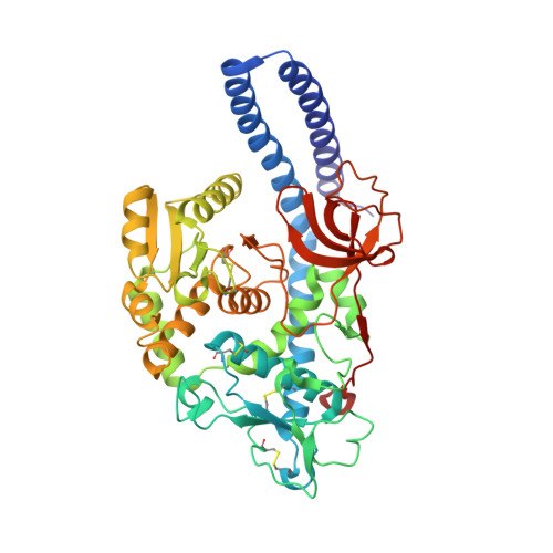
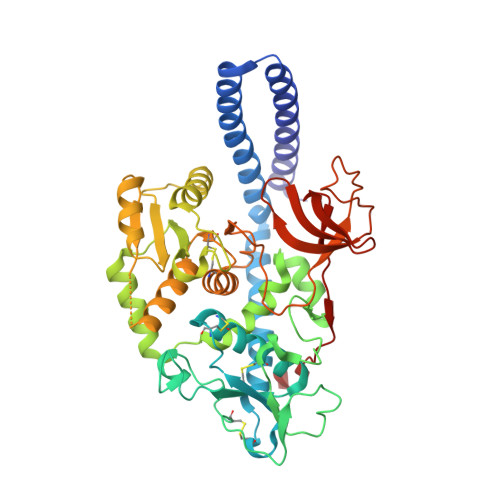
Structural basis for substrate specificity and catalysis of alpha 1,6-fucosyltransferase.

Garcia-Garcia, A.Ceballos-Laita, L.Serna, S.Artschwager, R.Reichardt, N.C.Corzana, F.Hurtado-Guerrero, R.

(2020) Nat Commun 11: 973-973

  • DOI: https://doi.org/10.1038/s41467-020-14794-z
  • Primary Citation of Related Structures:  
    6TKV

  • PubMed Abstract: 

    Core-fucosylation is an essential biological modification by which a fucose is transferred from GDP-β-L-fucose to the innermost N-acetylglucosamine residue of N-linked glycans. A single human enzyme α1,6-fucosyltransferase (FUT8) is the only enzyme responsible for this modification via the addition of an α-1,6-linked fucose to N-glycans. To date, the details of substrate recognition and catalysis by FUT8 remain unknown. Here, we report the crystal structure of FUT8 complexed with GDP and a biantennary complex N-glycan (G0), which provides insight into both substrate recognition and catalysis. FUT8 follows an S N 2 mechanism and deploys a series of loops and an α-helix which all contribute in forming the binding site. An exosite, formed by one of these loops and an SH3 domain, is responsible for the recognition of branched sugars, making contacts specifically to the α1,3 arm GlcNAc, a feature required for catalysis. This information serves as a framework for inhibitor design, and helps to assess its potential as a therapeutic target.


  • Organizational Affiliation

    Institute of Biocomputation and Physics of Complex Systems (BIFI), University of Zaragoza, Mariano Esquillor s/n, Campus Rio Ebro, Edificio I+D, Zaragoza, Spain.


Macromolecules
Find similar proteins by:  (by identity cutoff)  |  3D Structure
Entity ID: 1
MoleculeChains Sequence LengthOrganismDetailsImage
Alpha-(1,6)-fucosyltransferase468Homo sapiensMutation(s): 0 
Gene Names: FUT8
EC: 2.4.1.68
UniProt & NIH Common Fund Data Resources
Find proteins for Q9BYC5 (Homo sapiens)
Explore Q9BYC5 
Go to UniProtKB:  Q9BYC5
PHAROS:  Q9BYC5
GTEx:  ENSG00000033170 
Entity Groups  
Sequence Clusters30% Identity50% Identity70% Identity90% Identity95% Identity100% Identity
UniProt GroupQ9BYC5
Sequence Annotations
Expand
  • Reference Sequence
Find similar proteins by:  (by identity cutoff)  |  3D Structure
Entity ID: 2
MoleculeChains Sequence LengthOrganismDetailsImage
Alpha-(1,6)-fucosyltransferase453Homo sapiensMutation(s): 0 
Gene Names: FUT8
EC: 2.4.1.68
UniProt & NIH Common Fund Data Resources
Find proteins for Q9BYC5 (Homo sapiens)
Explore Q9BYC5 
Go to UniProtKB:  Q9BYC5
PHAROS:  Q9BYC5
GTEx:  ENSG00000033170 
Entity Groups  
Sequence Clusters30% Identity50% Identity70% Identity90% Identity95% Identity100% Identity
UniProt GroupQ9BYC5
Sequence Annotations
Expand
  • Reference Sequence
Oligosaccharides

Help

Entity ID: 3
MoleculeChains Length2D Diagram Glycosylation3D Interactions
2-acetamido-2-deoxy-beta-D-glucopyranose-(1-2)-alpha-D-mannopyranose-(1-3)-[2-acetamido-2-deoxy-beta-D-glucopyranose-(1-2)-alpha-D-mannopyranose-(1-6)]beta-D-mannopyranose-(1-4)-2-acetamido-2-deoxy-beta-D-glucopyranose-(1-4)-2-acetamido-2-deoxy-beta-D-glucopyranose
C, D
7N/A
Glycosylation Resources
GlyTouCan:  G39213VZ
GlyCosmos:  G39213VZ
GlyGen:  G39213VZ
Small Molecules
Ligands 2 Unique
IDChains Name / Formula / InChI Key2D Diagram3D Interactions
GDP
Query on GDP

Download Ideal Coordinates CCD File 
E [auth A],
J [auth B]
GUANOSINE-5'-DIPHOSPHATE
C10 H15 N5 O11 P2
QGWNDRXFNXRZMB-UUOKFMHZSA-N
GOL
Query on GOL

Download Ideal Coordinates CCD File 
F [auth A]
G [auth A]
H [auth A]
I [auth A]
K [auth B]
F [auth A],
G [auth A],
H [auth A],
I [auth A],
K [auth B],
L [auth B],
M [auth B],
N [auth B],
O [auth B],
P [auth B],
Q [auth B],
R [auth B],
S [auth B],
T [auth B],
U [auth B],
V [auth B],
W [auth B]
GLYCEROL
C3 H8 O3
PEDCQBHIVMGVHV-UHFFFAOYSA-N
Experimental Data & Validation

Experimental Data

  • Method: X-RAY DIFFRACTION
  • Resolution: 1.95 Å
  • R-Value Free: 0.219 
  • R-Value Work: 0.185 
  • R-Value Observed: 0.187 
  • Space Group: C 1 2 1
Unit Cell:
Length ( Å )Angle ( ˚ )
a = 208.039α = 90
b = 68.589β = 149.9
c = 173.921γ = 90
Software Package:
Software NamePurpose
REFMACrefinement
XDSdata reduction
SCALAdata scaling
PHASERphasing

Structure Validation

View Full Validation Report


Ligand Structure Quality Assessment 


Entry History 

Deposition Data

Revision History  (Full details and data files)

  • Version 1.0: 2020-01-29
    Type: Initial release
  • Version 1.1: 2020-02-19
    Changes: Data collection, Database references
  • Version 1.2: 2020-03-04
    Changes: Database references
  • Version 2.0: 2020-07-29
    Type: Remediation
    Reason: Carbohydrate remediation
    Changes: Atomic model, Data collection, Derived calculations, Structure summary
  • Version 2.1: 2024-01-24
    Changes: Data collection, Database references, Derived calculations, Refinement description, Structure summary