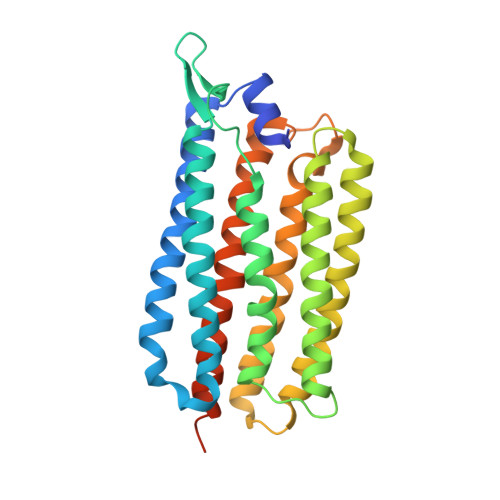
6TK2

Femtosecond to millisecond structural changes in a light-driven sodium pump: 1ms structure of KR2 with extrapolated, light and dark datasets


Experimental Data Snapshot

  • Method: X-RAY DIFFRACTION
  • Resolution: 2.50 Å
  • R-Value Free: 0.324 
  • R-Value Work: 0.258 
  • R-Value Observed: 0.263 

wwPDB Validation   3D Report Full Report


Ligand Structure Quality Assessment 


This is version 1.3 of the entry. See complete history


Literature

Femtosecond-to-millisecond structural changes in a light-driven sodium pump.

Skopintsev, P.Ehrenberg, D.Weinert, T.James, D.Kar, R.K.Johnson, P.J.M.Ozerov, D.Furrer, A.Martiel, I.Dworkowski, F.Nass, K.Knopp, G.Cirelli, C.Arrell, C.Gashi, D.Mous, S.Wranik, M.Gruhl, T.Kekilli, D.Brunle, S.Deupi, X.Schertler, G.F.X.Benoit, R.M.Panneels, V.Nogly, P.Schapiro, I.Milne, C.Heberle, J.Standfuss, J.

(2020) Nature 583: 314-318

  • DOI: https://doi.org/10.1038/s41586-020-2307-8
  • Primary Citation of Related Structures:  
    6TK1, 6TK2, 6TK3, 6TK4, 6TK5, 6TK6, 6TK7

  • PubMed Abstract: 

    Light-driven sodium pumps actively transport small cations across cellular membranes 1 . These pumps are used by microorganisms to convert light into membrane potential and have become useful optogenetic tools with applications in neuroscience. Although the resting state structures of the prototypical sodium pump Krokinobacter eikastus rhodopsin 2 (KR2) have been solved 2,3 , it is unclear how structural alterations over time allow sodium to be translocated against a concentration gradient. Here, using the Swiss X-ray Free Electron Laser 4 , we have collected serial crystallographic data at ten pump-probe delays from femtoseconds to milliseconds. High-resolution structural snapshots throughout the KR2 photocycle show how retinal isomerization is completed on the femtosecond timescale and changes the local structure of the binding pocket in the early nanoseconds. Subsequent rearrangements and deprotonation of the retinal Schiff base open an electrostatic gate in microseconds. Structural and spectroscopic data, in combination with quantum chemical calculations, indicate that a sodium ion binds transiently close to the retinal within one millisecond. In the last structural intermediate, at 20 milliseconds after activation, we identified a potential second sodium-binding site close to the extracellular exit. These results provide direct molecular insight into the dynamics of active cation transport across biological membranes.


  • Organizational Affiliation

    Laboratory of Biomolecular Research, Biology and Chemistry Division, Paul Scherrer Institut, Villigen, Switzerland.


Macromolecules
Find similar proteins by:  (by identity cutoff)  |  3D Structure
Entity ID: 1
MoleculeChains Sequence LengthOrganismDetailsImage
Sodium pumping rhodopsin290Dokdonia eikastaMutation(s): 0 
Gene Names: NaR
Membrane Entity: Yes 
UniProt
Find proteins for N0DKS8 (Dokdonia eikasta)
Explore N0DKS8 
Go to UniProtKB:  N0DKS8
Entity Groups  
Sequence Clusters30% Identity50% Identity70% Identity90% Identity95% Identity100% Identity
UniProt GroupN0DKS8
Sequence Annotations
Expand
  • Reference Sequence
Small Molecules
Ligands 3 Unique
IDChains Name / Formula / InChI Key2D Diagram3D Interactions
RET (Subject of Investigation/LOI)
Query on RET

Download Ideal Coordinates CCD File 
B [auth A]RETINAL
C20 H28 O
NCYCYZXNIZJOKI-OVSJKPMPSA-N
LFA
Query on LFA

Download Ideal Coordinates CCD File 
AA [auth A]
BA [auth A]
C [auth A]
D [auth A]
E [auth A]
AA [auth A],
BA [auth A],
C [auth A],
D [auth A],
E [auth A],
F [auth A],
G [auth A],
H [auth A],
I [auth A],
J [auth A],
K [auth A],
L [auth A],
M [auth A],
N [auth A],
O [auth A],
P [auth A],
Q [auth A],
R [auth A],
S [auth A],
T [auth A],
U [auth A],
V [auth A],
W [auth A],
X [auth A],
Y [auth A],
Z [auth A]
EICOSANE
C20 H42
CBFCDTFDPHXCNY-UHFFFAOYSA-N
NA (Subject of Investigation/LOI)
Query on NA

Download Ideal Coordinates CCD File 
CA [auth A]SODIUM ION
Na
FKNQFGJONOIPTF-UHFFFAOYSA-N
Experimental Data & Validation

Experimental Data

  • Method: X-RAY DIFFRACTION
  • Resolution: 2.50 Å
  • R-Value Free: 0.324 
  • R-Value Work: 0.258 
  • R-Value Observed: 0.263 
  • Space Group: I 2 2 2
Unit Cell:
Length ( Å )Angle ( ˚ )
a = 41.54α = 90
b = 84.48β = 90
c = 235.56γ = 90
Software Package:
Software NamePurpose
PHENIXrefinement
PDB_EXTRACTdata extraction
CrystFELdata reduction
CrystFELdata scaling
PHASERphasing

Structure Validation

View Full Validation Report


Ligand Structure Quality Assessment 


Entry History & Funding Information

Deposition Data


Funding OrganizationLocationGrant Number
Swiss National Science FoundationSwitzerland31003A_141235
Swiss National Science FoundationSwitzerland31003A_159558
Swiss National Science FoundationSwitzerlandPZ00P3_174169
German Research FoundationGermanySFB-1078, project B3
German Research FoundationGermanySPP-1926, HE 2063/6-1
European Research CouncilSwitzerlandMarie-Sklodowska-Curie No 701646
European Research CouncilSwitzerlandMarie-Sklodowska-Curie No 701647
European Research CouncilIsrael678169
German Research FoundationIsraelSFB 1078 Protonation Dynamics in Protein Function Mercator fellowship

Revision History  (Full details and data files)

  • Version 1.0: 2020-05-27
    Type: Initial release
  • Version 1.1: 2020-06-17
    Changes: Database references
  • Version 1.2: 2020-07-22
    Changes: Database references
  • Version 1.3: 2024-01-24
    Changes: Data collection, Database references, Derived calculations, Refinement description