6TEL | pdb_00006tel

Crystal structure of Dot1L in complex with an inhibitor (compound 10).


Experimental Data Snapshot

  • Method: X-RAY DIFFRACTION
  • Resolution: 2.19 Å
  • R-Value Free: 
    0.212 (Depositor), 0.215 (DCC) 
  • R-Value Work: 
    0.185 (Depositor), 0.185 (DCC) 
  • R-Value Observed: 
    0.187 (Depositor) 

Starting Model: experimental
View more details

wwPDB Validation 3D Report Full Report

Validation slider image for 6TEL

Ligand Structure Quality Assessment 


This is version 1.2 of the entry. See complete history

Literature

New Potent DOT1L Inhibitors forin VivoEvaluation in Mouse.

Stauffer, F.Weiss, A.Scheufler, C.Mobitz, H.Ragot, C.Beyer, K.S.Calkins, K.Guthy, D.Kiffe, M.Van Eerdenbrugh, B.Tiedt, R.Gaul, C.

(2019) ACS Med Chem Lett 10: 1655-1660

  • DOI: https://doi.org/10.1021/acsmedchemlett.9b00452
  • Primary Citation Related Structures: 
    6TE6, 6TEL, 6TEN

  • PubMed Abstract: 

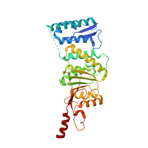
    In MLL-rearranged cancer cells, disruptor of telomeric silencing 1-like protein (DOT1L) is aberrantly recruited to ectopic loci leading to local hypermethylation of H3K79 and consequently misexpression of leukemogenic genes. A structure-guided optimization of a HTS hit led to the discovery of DOT1L inhibitors with subnanomolar potency, allowing testing of the therapeutic principle of DOT1L inhibition in a preclinical mouse tumor xenograft model. Compounds displaying good exposure in mouse and nanomolar inhibition of target gene expression in cells were obtained and tested in vivo.


  • Organizational Affiliation
    • Novartis Institutes for Biomedical Research, 4056 Basel, Switzerland.

Macromolecule Content 

  • Total Structure Weight: 78.27 kDa 
  • Atom Count: 5,314 
  • Modeled Residue Count: 628 
  • Deposited Residue Count: 668 
  • Unique protein chains: 1

Macromolecules

Find similar proteins by:|  3D Structure
Entity ID: 1
MoleculeChains  Sequence LengthOrganismDetailsImage
Histone-lysine N-methyltransferase, H3 lysine-79 specific
A, B
334Homo sapiensMutation(s): 0 
EC: 2.1.1.43 (PDB Primary Data), 2.1.1.360 (UniProt)
UniProt & NIH Common Fund Data Resources
Find proteins for Q8TEK3 (Homo sapiens)
Explore Q8TEK3 
Go to UniProtKB:  Q8TEK3
PHAROS:  Q8TEK3
GTEx:  ENSG00000104885 
Entity Groups
Sequence Clusters30% Identity50% Identity70% Identity90% Identity95% Identity100% Identity
UniProt GroupQ8TEK3
Sequence Annotations
Expand
Reference Sequence

Small Molecules

Ligands 2 Unique
IDChains Name / Formula / InChI Key2D Diagram3D Interactions
N4Z
(Subject of Investigation/LOI)

Query on N4Z


Download:Ideal Coordinates CCD File
C [auth A],
D [auth B]
~{N}1-[(~{S})-[2,2-bis(fluoranyl)-1,3-benzodioxol-4-yl]-(3-chloranylpyridin-2-yl)methyl]-~{N}2-(4-methoxy-6-piperazin-1-yl-1,3,5-triazin-2-yl)-4-methylsulfonyl-benzene-1,2-diamine
C28 H27 Cl F2 N8 O5 S
QJIMSJUUARCROQ-QFIPXVFZSA-N
K

Query on K


Download:Ideal Coordinates CCD File
E [auth B]POTASSIUM ION
K
NPYPAHLBTDXSSS-UHFFFAOYSA-N
Binding Affinity Annotations 
IDSourceBinding Affinity
N4Z BindingDB:  6TEL IC50: 0.11 (nM) from 1 assay(s)

Experimental Data & Validation

Experimental Data

  • Method: X-RAY DIFFRACTION
  • Resolution: 2.19 Å
  • R-Value Free:  0.212 (Depositor), 0.215 (DCC) 
  • R-Value Work:  0.185 (Depositor), 0.185 (DCC) 
  • R-Value Observed: 0.187 (Depositor) 
Space Group: P 63
Unit Cell:
Length ( Å )Angle ( ˚ )
a = 159.16α = 90
b = 159.16β = 90
c = 74.14γ = 120
Software Package:
Software NamePurpose
XDSdata reduction
XSCALEdata scaling
PHASERphasing
BUSTERrefinement
PDB_EXTRACTdata extraction

Structure Validation

View Full Validation Report


Ligand Structure Quality Assessment 


Entry History 

Deposition Data

Revision History  (Full details and data files)

  • Version 1.0: 2019-12-11
    Type: Initial release
  • Version 1.1: 2020-01-01
    Changes: Database references
  • Version 1.2: 2024-01-24
    Changes: Data collection, Database references, Derived calculations, Refinement description