6TE0 | pdb_00006te0

Cryo-EM structure of Euglena gracilis mitochondrial ATP synthase, OSCP/F1/c-ring, rotational state 3


Experimental Data Snapshot

  • Method: ELECTRON MICROSCOPY
  • Resolution: 3.92 Å
  • Aggregation State: PARTICLE 
  • Reconstruction Method: SINGLE PARTICLE 

wwPDB Validation 3D Report Full Report

Validation slider image for 6TE0

This is version 1.1 of the entry. See complete history

Literature

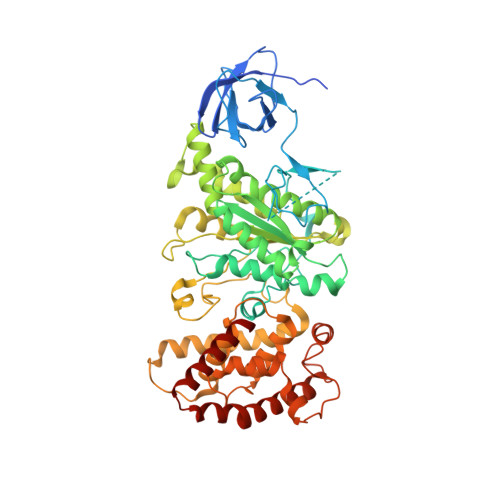
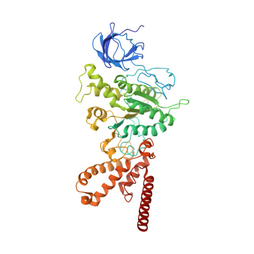
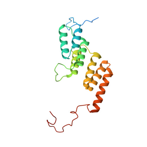
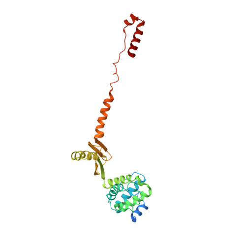
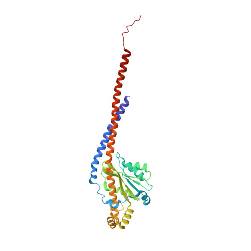
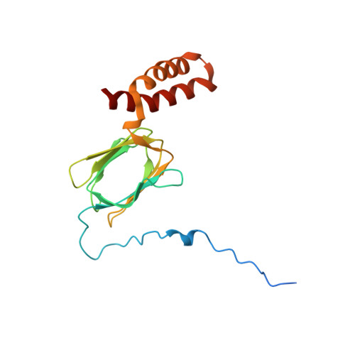
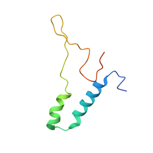
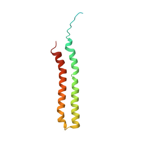
Structure of a mitochondrial ATP synthase with bound native cardiolipin.

Muhleip, A.McComas, S.E.Amunts, A.

(2019) Elife 8

  • DOI: https://doi.org/10.7554/eLife.51179
  • Primary Citation Related Structures: 
    6TDU, 6TDV, 6TDW, 6TDX, 6TDY, 6TDZ, 6TE0

  • PubMed Abstract: 

    The mitochondrial ATP synthase fuels eukaryotic cells with chemical energy. Here we report the cryo-EM structure of a divergent ATP synthase dimer from mitochondria of Euglena gracilis , a member of the phylum Euglenozoa that also includes human parasites. It features 29 different subunits, 8 of which are newly identified. The membrane region was determined to 2.8 Å resolution, enabling the identification of 37 associated lipids, including 25 cardiolipins, which provides insight into protein-lipid interactions and their functional roles. The rotor-stator interface comprises four membrane-embedded horizontal helices, including a distinct subunit a . The dimer interface is formed entirely by phylum-specific components, and a peripherally associated subcomplex contributes to the membrane curvature. The central and peripheral stalks directly interact with each other. Last, the ATPase inhibitory factor 1 (IF 1 ) binds in a mode that is different from human, but conserved in Trypanosomatids.


  • Organizational Affiliation
    • Science for Life Laboratory, Department of Biochemistry and Biophysics, Stockholm University, Solna, Sweden.

Macromolecule Content 

  • Total Structure Weight: 612.44 kDa 
  • Atom Count: 39,353 
  • Modeled Residue Count: 5,136 
  • Deposited Residue Count: 5,606 
  • Unique protein chains: 8

Macromolecules

Find similar proteins by:|  3D Structure
Entity ID: 1
MoleculeChains  Sequence LengthOrganismDetailsImage
ATP synthase subunit alphaA [auth C],
B [auth A],
C [auth B]
561Euglena gracilisMutation(s): 0 
Find similar proteins by:|  3D Structure
Entity ID: 2
MoleculeChains  Sequence LengthOrganismDetailsImage
ATP synthase subunit betaD [auth F],
E [auth D],
F [auth E]
494Euglena gracilisMutation(s): 0 
Find similar proteins by:|  3D Structure
Entity ID: 3
MoleculeChains  Sequence LengthOrganismDetailsImage
Ribonucleoprotein P18G [auth L],
H [auth J],
I [auth K]
192Euglena gracilisMutation(s): 0 
Find similar proteins by:|  3D Structure
Entity ID: 4
MoleculeChains  Sequence LengthOrganismDetailsImage
oligomycin sensitivity conferring protein (OSCP)J [auth M]267Euglena gracilisMutation(s): 0 
Find similar proteins by:|  3D Structure
Entity ID: 5
MoleculeChains  Sequence LengthOrganismDetailsImage
ATP synthase F1 subunit gamma proteinK [auth G]306Euglena gracilisMutation(s): 0 
Find similar proteins by:|  3D Structure
Entity ID: 6
MoleculeChains  Sequence LengthOrganismDetailsImage
ATP synthase subunit deltaL [auth H]176Euglena gracilisMutation(s): 0 
Find similar proteins by:|  3D Structure
Entity ID: 7
MoleculeChains  Sequence LengthOrganismDetailsImage
ATP synthase subunit epsilonM [auth I]76Euglena gracilisMutation(s): 0 
Find similar proteins by:|  3D Structure
Entity ID: 8
MoleculeChains  Sequence LengthOrganismDetailsImage
ATP synthase subunit c104Euglena gracilisMutation(s): 0 

Small Molecules

Ligands 4 Unique
IDChains Name / Formula / InChI Key2D Diagram3D Interactions
ATP
(Subject of Investigation/LOI)

Query on ATP


Download:Ideal Coordinates CCD File
BA [auth B],
GA [auth E],
X [auth C],
Z [auth A]
ADENOSINE-5'-TRIPHOSPHATE
C10 H16 N5 O13 P3
ZKHQWZAMYRWXGA-KQYNXXCUSA-N
ADP
(Subject of Investigation/LOI)

Query on ADP


Download:Ideal Coordinates CCD File
DA [auth F]ADENOSINE-5'-DIPHOSPHATE
C10 H15 N5 O10 P2
XTWYTFMLZFPYCI-KQYNXXCUSA-N
TRT

Query on TRT


Download:Ideal Coordinates CCD File
FA [auth D]FRAGMENT OF TRITON X-100
C21 H36 O4
HEUDUECKTWTQQR-UHFFFAOYSA-N
MG

Query on MG


Download:Ideal Coordinates CCD File
AA [auth A],
CA [auth B],
EA [auth F],
HA [auth E],
Y [auth C]
MAGNESIUM ION
Mg
JLVVSXFLKOJNIY-UHFFFAOYSA-N

Experimental Data & Validation

Experimental Data

  • Method: ELECTRON MICROSCOPY
  • Resolution: 3.92 Å
  • Aggregation State: PARTICLE 
  • Reconstruction Method: SINGLE PARTICLE 
EM Software:
TaskSoftware PackageVersion
RECONSTRUCTIONRELION

Structure Validation

View Full Validation Report


Entry History 

Deposition Data

Revision History  (Full details and data files)

  • Version 1.0: 2019-11-27
    Type: Initial release
  • Version 1.1: 2024-05-22
    Changes: Data collection, Database references