6T9C

Crystal structure of the complex between PPARgamma LBD and the ligand NV1346 (3a)


Experimental Data Snapshot

  • Method: X-RAY DIFFRACTION
  • Resolution: 1.95 Å
  • R-Value Free: 0.262 
  • R-Value Work: 0.230 
  • R-Value Observed: 0.232 

wwPDB Validation   3D Report Full Report


Ligand Structure Quality Assessment 


This is version 1.3 of the entry. See complete history


Literature

Insights into PPAR gamma Phosphorylation and Its Inhibition Mechanism.

Montanari, R.Capelli, D.Yamamoto, K.Awaishima, H.Nishikata, K.Barendregt, A.Heck, A.J.R.Loiodice, F.Altieri, F.Paiardini, A.Grottesi, A.Pirone, L.Pedone, E.Peiretti, F.Brunel, J.M.Itoh, T.Pochetti, G.

(2020) J Med Chem 63: 4811-4823

  • DOI: https://doi.org/10.1021/acs.jmedchem.0c00048
  • Primary Citation of Related Structures:  
    6T9C

  • PubMed Abstract: 

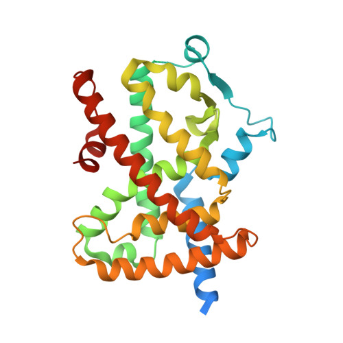
    PPARγ represents a key target for the treatment of type 2 diabetes and metabolic syndrome. Synthetic antidiabetic drugs activating PPARγ are accompanied by serious undesirable side effects related to their agonism. In the search for new PPARγ regulators, inhibitors of PPARγ phosphorylation on S245 mediated by CDK5 represent an opportunity for the development of an improved generation of antidiabetic drugs acting through this nuclear receptor. We have employed a multidisciplinary approach, including protein-protein docking, X-ray crystallography, NMR, HDX, MD simulations, and site-directed mutagenesis to investigate conformational changes in PPARγ that impair the ability of CDK5 to interact with PPARγ and hence inhibit PPARγ phosphorylation. Finally, we describe an alternative inhibition mechanism adopted by a ligand bound far from the phosphorylation site.


  • Organizational Affiliation

    Istituto di Cristallografia, Consiglio Nazionale delle Ricerche, Via Salaria km. 29.300, 00015 Monterotondo Stazione, Rome, Italy.


Macromolecules
Find similar proteins by:  (by identity cutoff)  |  3D Structure
Entity ID: 1
MoleculeChains Sequence LengthOrganismDetailsImage
Peroxisome proliferator-activated receptor gamma
A, B
304Homo sapiensMutation(s): 0 
Gene Names: PPARGNR1C3
UniProt & NIH Common Fund Data Resources
Find proteins for P37231 (Homo sapiens)
Explore P37231 
Go to UniProtKB:  P37231
PHAROS:  P37231
GTEx:  ENSG00000132170 
Entity Groups  
Sequence Clusters30% Identity50% Identity70% Identity90% Identity95% Identity100% Identity
UniProt GroupP37231
Sequence Annotations
Expand
  • Reference Sequence
Small Molecules
Ligands 1 Unique
IDChains Name / Formula / InChI Key2D Diagram3D Interactions
MX8 (Subject of Investigation/LOI)
Query on MX8

Download Ideal Coordinates CCD File 
C [auth A]4-hexoxy-~{N}-[(2~{S})-3-methyl-1-(oxidanylamino)-1-oxidanylidene-butan-2-yl]benzamide
C18 H28 N2 O4
GUGHZGUQZCBZHP-INIZCTEOSA-N
Experimental Data & Validation

Experimental Data

  • Method: X-RAY DIFFRACTION
  • Resolution: 1.95 Å
  • R-Value Free: 0.262 
  • R-Value Work: 0.230 
  • R-Value Observed: 0.232 
  • Space Group: C 1 2 1
Unit Cell:
Length ( Å )Angle ( ˚ )
a = 93.78α = 90
b = 60.98β = 103.5
c = 119γ = 90
Software Package:
Software NamePurpose
PHENIXrefinement
PDB_EXTRACTdata extraction
MOSFLMdata reduction
SCALAdata scaling
AMoREphasing

Structure Validation

View Full Validation Report


Ligand Structure Quality Assessment 


Entry History 

Deposition Data

Revision History  (Full details and data files)

  • Version 1.0: 2020-02-26
    Type: Initial release
  • Version 1.1: 2020-06-03
    Changes: Database references
  • Version 1.2: 2020-06-10
    Changes: Database references
  • Version 1.3: 2024-01-24
    Changes: Data collection, Database references, Refinement description