6T8Z

NAD+-dependent fungal formate dehydrogenase from Chaetomium thermophilum: A ternary complex with the oxidised form of the cofactor NAD+ and the substrate formate both at a primary and secondary sites.


Experimental Data Snapshot

  • Method: X-RAY DIFFRACTION
  • Resolution: 1.21 Å
  • R-Value Free: 0.211 
  • R-Value Work: 0.188 

wwPDB Validation   3D Report Full Report


Ligand Structure Quality Assessment 


This is version 1.2 of the entry. See complete history


Literature

Structural insights into the NAD + -dependent formate dehydrogenase mechanism revealed from the NADH complex and the formate NAD + ternary complex of the Chaetomium thermophilum enzyme.

Yilmazer, B.Isupov, M.N.De Rose, S.A.Bulut, H.Benninghoff, J.C.Binay, B.Littlechild, J.A.

(2020) J Struct Biol 212: 107657-107657

  • DOI: https://doi.org/10.1016/j.jsb.2020.107657
  • Primary Citation of Related Structures:  
    6T8Y, 6T8Z, 6T92, 6T94

  • PubMed Abstract: 

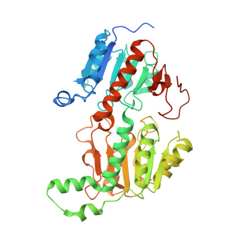
    The removal of carbon dioxide from the waste streams of industrial processes is a major challenge for creation of a sustainable circular economy. This makes the synthesis of formate from CO 2 by NAD + dependent formate dehydrogenases (FDHs) an attractive process for this purpose. The efficiency of this reaction is however low and to achieve a viable industrial process an optimised engineered enzyme needs to be developed. In order to understand the detailed enzymatic mechanism of catalysis structures of different cofactor and substrate complexes of the FDH from the thermophilic filamentous fungus, Chaetomium thermophilum have been determined to 1.2-1.3 Å resolution. The substrate formate is shown to be held by four hydrogen bonds in the FDH catalytic site within the ternary complex with substrate and NAD + and a secondary formate binding site is observed in crystals soaked with substrate. Water molecules are excluded from the FDH catalytic site when the substrate is bound. The angle between the plane of the NAD + cofactor pyridine ring and the plane of the formate molecule is around 27°. Additionally, structures of a FDH mutant enzyme, N120C, in complex with the reduced form of the cofactor have also been determined both in the presence and absence of formate bound at the secondary site. These structures provide further understanding of the catalytic mechanism of this fungal enzyme.


  • Organizational Affiliation

    Department of Molecular Biology and Genetics, Gebze Technical University, 41400 Gebze, Kocaeli, Turkey.


Macromolecules
Find similar proteins by:  (by identity cutoff)  |  3D Structure
Entity ID: 1
MoleculeChains Sequence LengthOrganismDetailsImage
Formate dehydrogenaseA [auth AAA],
B [auth BBB],
C [auth CCC],
D [auth DDD]
410Thermochaetoides thermophila DSM 1495Mutation(s): 0 
Gene Names: FDHCTHT_0067590
EC: 1.17.1.9
UniProt
Find proteins for G0SGU4 (Chaetomium thermophilum (strain DSM 1495 / CBS 144.50 / IMI 039719))
Explore G0SGU4 
Go to UniProtKB:  G0SGU4
Entity Groups  
Sequence Clusters30% Identity50% Identity70% Identity90% Identity95% Identity100% Identity
UniProt GroupG0SGU4
Sequence Annotations
Expand
  • Reference Sequence
Small Molecules
Ligands 4 Unique
IDChains Name / Formula / InChI Key2D Diagram3D Interactions
NAD (Subject of Investigation/LOI)
Query on NAD

Download Ideal Coordinates CCD File 
DA [auth DDD],
I [auth AAA],
R [auth BBB],
X [auth CCC]
NICOTINAMIDE-ADENINE-DINUCLEOTIDE
C21 H27 N7 O14 P2
BAWFJGJZGIEFAR-NNYOXOHSSA-N
PEG
Query on PEG

Download Ideal Coordinates CCD File 
AA [auth DDD]
E [auth AAA]
F [auth AAA]
O [auth BBB]
T [auth CCC]
AA [auth DDD],
E [auth AAA],
F [auth AAA],
O [auth BBB],
T [auth CCC],
U [auth CCC],
Z [auth DDD]
DI(HYDROXYETHYL)ETHER
C4 H10 O3
MTHSVFCYNBDYFN-UHFFFAOYSA-N
EDO
Query on EDO

Download Ideal Coordinates CCD File 
J [auth AAA]
K [auth AAA]
L [auth AAA]
M [auth AAA]
N [auth AAA]
J [auth AAA],
K [auth AAA],
L [auth AAA],
M [auth AAA],
N [auth AAA],
S [auth BBB],
Y [auth CCC]
1,2-ETHANEDIOL
C2 H6 O2
LYCAIKOWRPUZTN-UHFFFAOYSA-N
FMT (Subject of Investigation/LOI)
Query on FMT

Download Ideal Coordinates CCD File 
BA [auth DDD]
CA [auth DDD]
G [auth AAA]
H [auth AAA]
P [auth BBB]
BA [auth DDD],
CA [auth DDD],
G [auth AAA],
H [auth AAA],
P [auth BBB],
Q [auth BBB],
V [auth CCC],
W [auth CCC]
FORMIC ACID
C H2 O2
BDAGIHXWWSANSR-UHFFFAOYSA-N
Experimental Data & Validation

Experimental Data

  • Method: X-RAY DIFFRACTION
  • Resolution: 1.21 Å
  • R-Value Free: 0.211 
  • R-Value Work: 0.188 
  • Space Group: P 1
Unit Cell:
Length ( Å )Angle ( ˚ )
a = 50.395α = 85.689
b = 94.515β = 89.927
c = 94.33γ = 81.939
Software Package:
Software NamePurpose
REFMACrefinement
XDSdata reduction
XDSdata scaling
MoRDaphasing

Structure Validation

View Full Validation Report


Ligand Structure Quality Assessment 


Entry History 

Deposition Data

Revision History  (Full details and data files)

  • Version 1.0: 2020-11-18
    Type: Initial release
  • Version 1.1: 2021-06-02
    Changes: Database references
  • Version 1.2: 2024-01-24
    Changes: Data collection, Database references, Derived calculations, Refinement description