6T85

Urocanate reductase in complex with ADP


Experimental Data Snapshot

  • Method: X-RAY DIFFRACTION
  • Resolution: 1.10 Å
  • R-Value Free: 0.130 
  • R-Value Work: 0.107 
  • R-Value Observed: 0.109 

wwPDB Validation   3D Report Full Report


Ligand Structure Quality Assessment 


This is version 1.2 of the entry. See complete history


Literature

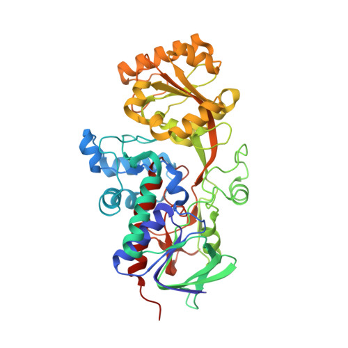
Structural characterization of the microbial enzyme urocanate reductase mediating imidazole propionate production.

Venskutonyte, R.Koh, A.Stenstrom, O.Khan, M.T.Lundqvist, A.Akke, M.Backhed, F.Lindkvist-Petersson, K.

(2021) Nat Commun 12: 1347-1347

  • DOI: https://doi.org/10.1038/s41467-021-21548-y
  • Primary Citation of Related Structures:  
    6T85, 6T86, 6T87, 6T88

  • PubMed Abstract: 

    The human microbiome can produce metabolites that modulate insulin signaling. Type 2 diabetes patients have increased circulating concentrations of the microbially produced histidine metabolite, imidazole propionate (ImP) and administration of ImP in mice resulted in impaired glucose tolerance. Interestingly, the fecal microbiota of the patients had increased capacity to produce ImP, which is mediated by the bacterial enzyme urocanate reductase (UrdA). Here, we describe the X-ray structures of the ligand-binding domains of UrdA in four different states, representing the structural transitions along the catalytic reaction pathway of this unexplored enzyme linked to disease in humans. The structures in combination with functional data provide key insights into the mechanism of action of UrdA that open new possibilities for drug development strategies targeting type 2 diabetes.


  • Organizational Affiliation

    Experimental Medical Science, Medical Structural Biology, BMC C13, Lund University, Lund, Sweden.


Macromolecules
Find similar proteins by:  (by identity cutoff)  |  3D Structure
Entity ID: 1
MoleculeChains Sequence LengthOrganismDetailsImage
Urocanate reductase460Shewanella oneidensis MR-1Mutation(s): 0 
Gene Names: urdASO_4620
EC: 1.3.99.33
UniProt
Find proteins for Q8CVD0 (Shewanella oneidensis (strain MR-1))
Explore Q8CVD0 
Go to UniProtKB:  Q8CVD0
Entity Groups  
Sequence Clusters30% Identity50% Identity70% Identity90% Identity95% Identity100% Identity
UniProt GroupQ8CVD0
Sequence Annotations
Expand
  • Reference Sequence
Small Molecules
Ligands 3 Unique
IDChains Name / Formula / InChI Key2D Diagram3D Interactions
ADP
Query on ADP

Download Ideal Coordinates CCD File 
B [auth A]ADENOSINE-5'-DIPHOSPHATE
C10 H15 N5 O10 P2
XTWYTFMLZFPYCI-KQYNXXCUSA-N
GOL
Query on GOL

Download Ideal Coordinates CCD File 
H [auth A],
I [auth A],
J [auth A]
GLYCEROL
C3 H8 O3
PEDCQBHIVMGVHV-UHFFFAOYSA-N
CL
Query on CL

Download Ideal Coordinates CCD File 
C [auth A],
D [auth A],
E [auth A],
F [auth A],
G [auth A]
CHLORIDE ION
Cl
VEXZGXHMUGYJMC-UHFFFAOYSA-M
Experimental Data & Validation

Experimental Data

  • Method: X-RAY DIFFRACTION
  • Resolution: 1.10 Å
  • R-Value Free: 0.130 
  • R-Value Work: 0.107 
  • R-Value Observed: 0.109 
  • Space Group: P 1 21 1
Unit Cell:
Length ( Å )Angle ( ˚ )
a = 41.839α = 90
b = 95.762β = 91.04
c = 63.2γ = 90
Software Package:
Software NamePurpose
XDSdata reduction
Aimlessdata scaling
PHASERphasing
PHENIXrefinement
PDB_EXTRACTdata extraction

Structure Validation

View Full Validation Report


Ligand Structure Quality Assessment 


Entry History & Funding Information

Deposition Data


Funding OrganizationLocationGrant Number
Swedish Research CouncilSweden2016-01319
Swedish Research CouncilSweden2017-05816
European Research CouncilSweden780659

Revision History  (Full details and data files)

  • Version 1.0: 2021-03-03
    Type: Initial release
  • Version 1.1: 2021-03-10
    Changes: Database references
  • Version 1.2: 2024-01-24
    Changes: Data collection, Database references, Refinement description