6T7G

Bacteroides salyersiae GH164 beta-mannosidase in complex with mannoimidazole


Experimental Data Snapshot

  • Method: X-RAY DIFFRACTION
  • Resolution: 1.80 Å
  • R-Value Free: 0.231 
  • R-Value Work: 0.206 

wwPDB Validation   3D Report Full Report


Ligand Structure Quality Assessment 


This is version 1.3 of the entry. See complete history


Literature

Structure and function ofBs164 beta-mannosidase fromBacteroides salyersiaethe founding member of glycoside hydrolase family GH164.

Armstrong, Z.Davies, G.J.

(2020) J Biol Chem 295: 4316-4326

  • DOI: https://doi.org/10.1074/jbc.RA119.011591
  • Primary Citation of Related Structures:  
    6T5O, 6T6G, 6T75, 6T7G

  • PubMed Abstract: 

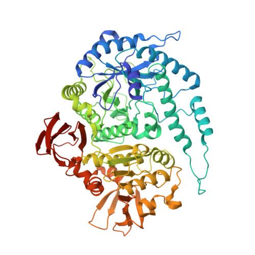
    Recent work exploring protein sequence space has revealed a new glycoside hydrolase (GH) family (GH164) of putative mannosidases. GH164 genes are present in several commensal bacteria, implicating these genes in the degradation of dietary glycans. However, little is known about the structure, mechanism of action, and substrate specificity of these enzymes. Herein we report the biochemical characterization and crystal structures of the founding member of this family ( Bs 164) from the human gut symbiont Bacteroides salyersiae. Previous reports of this enzyme indicated that it has α-mannosidase activity, however, we conclusively show that it cleaves only β-mannose linkages. Using NMR spectroscopy, detailed enzyme kinetics of WT and mutant Bs 164, and multiangle light scattering we found that it is a trimeric retaining β-mannosidase, that is susceptible to several known mannosidase inhibitors. X-ray crystallography revealed the structure of Bs 164, the first known structure of a GH164, at 1.91 Å resolution. Bs 164 is composed of three domains: a (β/α) 8 barrel, a trimerization domain, and a β-sandwich domain, representing a previously unobserved structural-fold for β-mannosidases. Structures of Bs 164 at 1.80-2.55 Å resolution in complex with the inhibitors noeuromycin, mannoimidazole, or 2,4-dinitrophenol 2-deoxy-2-fluoro-mannoside reveal the residues essential for specificity and catalysis including the catalytic nucleophile (Glu-297) and acid/base residue (Glu-160). These findings further our knowledge of the mechanisms commensal microbes use for nutrient acquisition.


  • Organizational Affiliation

    Department of Chemistry, Structural Biology Laboratory, The University of York, York YO10 5DD, United Kingdom.


Macromolecules
Find similar proteins by:  (by identity cutoff)  |  3D Structure
Entity ID: 1
MoleculeChains Sequence LengthOrganismDetailsImage
Glyco_hydro_42M domain-containing protein674Bacteroides salyersiae CL02T12C01Mutation(s): 0 
Gene Names: HMPREF1071_03408
UniProt
Find proteins for I9SUA3 (Bacteroides salyersiae CL02T12C01)
Explore I9SUA3 
Go to UniProtKB:  I9SUA3
Entity Groups  
Sequence Clusters30% Identity50% Identity70% Identity90% Identity95% Identity100% Identity
UniProt GroupI9SUA3
Sequence Annotations
Expand
  • Reference Sequence
Small Molecules
Ligands 3 Unique
IDChains Name / Formula / InChI Key2D Diagram3D Interactions
MVL (Subject of Investigation/LOI)
Query on MVL

Download Ideal Coordinates CCD File 
CA [auth FFF]
GA [auth EEE]
H [auth AAA]
L [auth DDD]
R [auth CCC]
CA [auth FFF],
GA [auth EEE],
H [auth AAA],
L [auth DDD],
R [auth CCC],
W [auth BBB]
(5R,6R,7S,8R)-5-(HYDROXYMETHYL)-5,6,7,8-TETRAHYDROIMIDAZO[1,2-A]PYRIDINE-6,7,8-TRIOL
C8 H12 N2 O4
RZRDQZQPTISYKY-JWXFUTCRSA-N
EDO
Query on EDO

Download Ideal Coordinates CCD File 
AA [auth FFF]
BA [auth FFF]
EA [auth EEE]
FA [auth EEE]
G [auth AAA]
AA [auth FFF],
BA [auth FFF],
EA [auth EEE],
FA [auth EEE],
G [auth AAA],
I [auth AAA],
K [auth DDD],
M [auth DDD],
O [auth CCC],
P [auth CCC],
Q [auth CCC],
T [auth BBB],
U [auth BBB],
V [auth BBB],
X [auth BBB],
Z [auth FFF]
1,2-ETHANEDIOL
C2 H6 O2
LYCAIKOWRPUZTN-UHFFFAOYSA-N
CL
Query on CL

Download Ideal Coordinates CCD File 
DA [auth FFF]
HA [auth EEE]
J [auth AAA]
N [auth DDD]
S [auth CCC]
DA [auth FFF],
HA [auth EEE],
J [auth AAA],
N [auth DDD],
S [auth CCC],
Y [auth BBB]
CHLORIDE ION
Cl
VEXZGXHMUGYJMC-UHFFFAOYSA-M
Experimental Data & Validation

Experimental Data

  • Method: X-RAY DIFFRACTION
  • Resolution: 1.80 Å
  • R-Value Free: 0.231 
  • R-Value Work: 0.206 
  • Space Group: P 1
Unit Cell:
Length ( Å )Angle ( ˚ )
a = 69.71α = 92.29
b = 104.743β = 97.273
c = 170.568γ = 106.301
Software Package:
Software NamePurpose
REFMACrefinement
xia2data reduction
DIALSdata scaling
PHASERphasing

Structure Validation

View Full Validation Report


Ligand Structure Quality Assessment 


Entry History & Funding Information

Deposition Data


Funding OrganizationLocationGrant Number
Biotechnology and Biological Sciences Research CouncilUnited KingdomBB/R001162/1
Biotechnology and Biological Sciences Research CouncilUnited KingdomBB/M011151/1

Revision History  (Full details and data files)

  • Version 1.0: 2019-12-25
    Type: Initial release
  • Version 1.1: 2020-01-08
    Changes: Database references
  • Version 1.2: 2020-04-08
    Changes: Database references
  • Version 1.3: 2024-01-24
    Changes: Data collection, Database references, Derived calculations, Refinement description