6SZN

Crystal structure of YTHDC1 with fragment 8 (DHU_DC1_006)


Experimental Data Snapshot

  • Method: X-RAY DIFFRACTION
  • Resolution: 1.47 Å
  • R-Value Free: 0.218 
  • R-Value Work: 0.183 
  • R-Value Observed: 0.184 

wwPDB Validation   3D Report Full Report


Ligand Structure Quality Assessment 


This is version 1.2 of the entry. See complete history


Literature

Selectively Disrupting m6A-Dependent Protein-RNA Interactions with Fragments.

Bedi, R.K.Huang, D.Wiedmer, L.Li, Y.Dolbois, A.Wojdyla, J.A.Sharpe, M.E.Caflisch, A.Sledz, P.

(2020) ACS Chem Biol 15: 618-625

  • DOI: https://doi.org/10.1021/acschembio.9b00894
  • Primary Citation of Related Structures:  
    6SYZ, 6SZ1, 6SZ2, 6SZ3, 6SZ7, 6SZ8, 6SZL, 6SZN, 6SZR, 6SZT, 6SZX, 6SZY, 6T01, 6T02, 6T03, 6T04, 6T05, 6T06, 6T07, 6T08, 6T09, 6T0A, 6T0C, 6T0D, 6T0O, 6T0X, 6T0Z, 6T10, 6T11, 6T12

  • PubMed Abstract: 

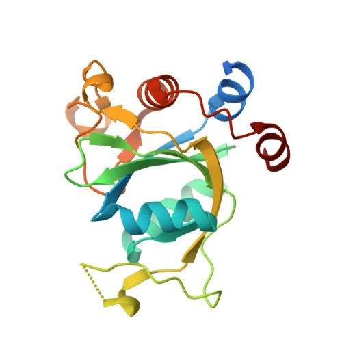
    We report a crystallographic analysis of small-molecule ligands of the human YTHDC1 domain that recognizes N6-methylated adenine (m 6 A) in RNA. The 30 binders are fragments (molecular weight < 300 g mol -1 ) that represent 10 different chemotypes identified by virtual screening. Despite the structural disorder of the binding site loop (residues 429-439), most of the 30 fragments emulate the two main interactions of the -NHCH 3 group of m 6 A. These interactions are the hydrogen bond to the backbone carbonyl of Ser378 and the van der Waals contacts with the tryptophan cage. Different chemical groups are involved in the conserved binding motifs. Some of the fragments show favorable ligand efficiency for YTHDC1 and selectivity against other m 6 A reader domains. The structural information is useful for the design of modulators of m 6 A recognition by YTHDC1.


  • Organizational Affiliation

    Department of Biochemistry, University of Zurich, Winterthurerstrasse 190, CH-8057 Zurich, Switzerland.


Macromolecules
Find similar proteins by:  (by identity cutoff)  |  3D Structure
Entity ID: 1
MoleculeChains Sequence LengthOrganismDetailsImage
YTH domain-containing protein 1
A, B
183Homo sapiensMutation(s): 0 
Gene Names: YTHDC1KIAA1966YT521
UniProt & NIH Common Fund Data Resources
Find proteins for Q96MU7 (Homo sapiens)
Explore Q96MU7 
Go to UniProtKB:  Q96MU7
PHAROS:  Q96MU7
GTEx:  ENSG00000083896 
Entity Groups  
Sequence Clusters30% Identity50% Identity70% Identity90% Identity95% Identity100% Identity
UniProt GroupQ96MU7
Sequence Annotations
Expand
  • Reference Sequence
Experimental Data & Validation

Experimental Data

  • Method: X-RAY DIFFRACTION
  • Resolution: 1.47 Å
  • R-Value Free: 0.218 
  • R-Value Work: 0.183 
  • R-Value Observed: 0.184 
  • Space Group: P 1 21 1
Unit Cell:
Length ( Å )Angle ( ˚ )
a = 39.83α = 90
b = 104.115β = 104.82
c = 42.115γ = 90
Software Package:
Software NamePurpose
XDSdata reduction
PHASERphasing
PHENIXrefinement

Structure Validation

View Full Validation Report


Ligand Structure Quality Assessment 


Entry History & Funding Information

Deposition Data


Funding OrganizationLocationGrant Number
Swiss National Science FoundationSwitzerlandCRSII5_180345

Revision History  (Full details and data files)

  • Version 1.0: 2020-03-04
    Type: Initial release
  • Version 1.1: 2020-04-01
    Changes: Database references
  • Version 1.2: 2024-01-24
    Changes: Data collection, Database references, Refinement description