6SXI | pdb_00006sxi

Antibody-anti-idiotype complex: AP33 Fab (hepatitis C virus E2 antibody) - B2.1A scFv (anti-idiotype)


Experimental Data Snapshot

  • Method: X-RAY DIFFRACTION
  • Resolution: 1.85 Å
  • R-Value Free: 
    0.203 (Depositor), 0.205 (DCC) 
  • R-Value Work: 
    0.156 (Depositor), 0.159 (DCC) 
  • R-Value Observed: 
    0.158 (Depositor) 

wwPDB Validation 3D Report Full Report

Validation slider image for 6SXI

This is version 1.2 of the entry. See complete history

Literature

Development of a structural epitope mimic: an idiotypic approach to HCV vaccine design.

Cowton, V.M.Owsianka, A.M.Fadda, V.Ortega-Prieto, A.M.Cole, S.J.Potter, J.A.Skelton, J.K.Jeffrey, N.Di Lorenzo, C.Dorner, M.Taylor, G.L.Patel, A.H.

(2021) NPJ Vaccines 6: 7-7

  • DOI: https://doi.org/10.1038/s41541-020-00269-1
  • Primary Citation Related Structures: 
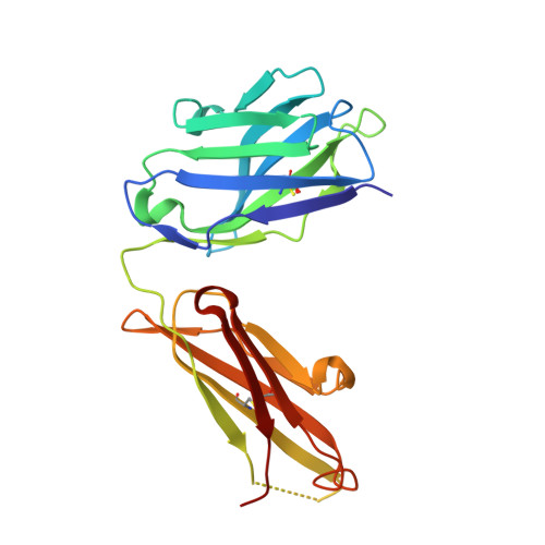
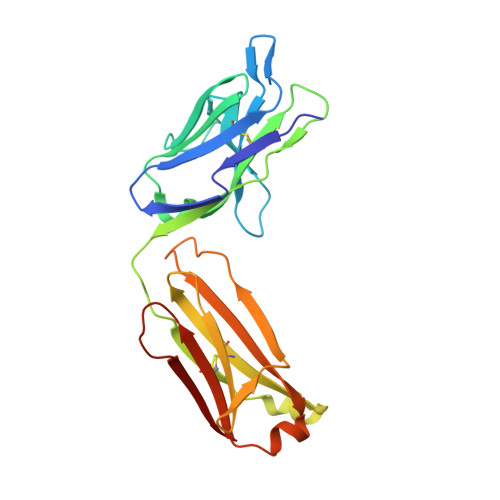
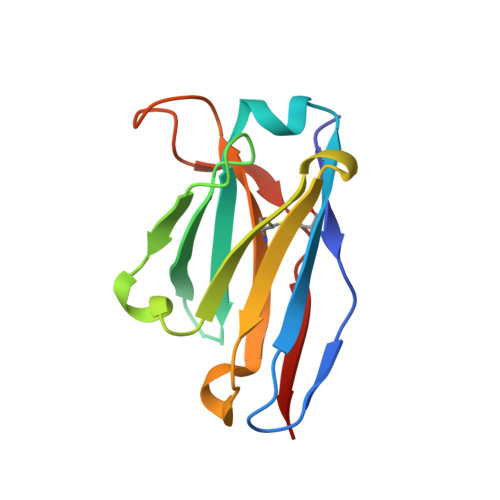
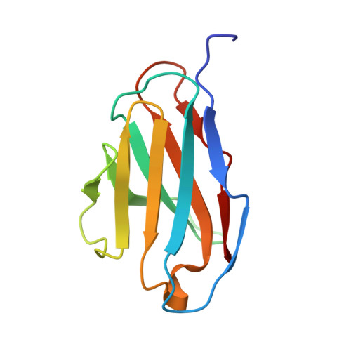
    6SXI

  • PubMed Abstract: 

    HCV vaccine development is stymied by the high genetic diversity of the virus and the variability of the envelope glycoproteins. One strategy to overcome this is to identify conserved, functionally important regions-such as the epitopes of broadly neutralizing antibodies (bNAbs)-and use these as a basis for structure-based vaccine design. Here, we report an anti-idiotype approach that has generated an antibody that mimics a highly conserved neutralizing epitope on HCV E2. Crucially, a mutagenesis screen was used to identify the antibody, designated B2.1 A, whose binding characteristics to the bNAb AP33 closely resemble those of the original antigen. Protein crystallography confirmed that B2.1 A is a structural mimic of the AP33 epitope. When used as an immunogen B2.1 A induced antibodies that recognized the same epitope and E2 residues as AP33 and most importantly protected against HCV challenge in a mouse model.


  • Organizational Affiliation
    • MRC-University of Glasgow Centre for Virus Research, Garscube Campus, 464 Bearsden Road, Glasgow, UK.

Macromolecule Content 

  • Total Structure Weight: 72.83 kDa 
  • Atom Count: 5,639 
  • Modeled Residue Count: 654 
  • Deposited Residue Count: 663 
  • Unique protein chains: 4

Macromolecules

Find similar proteins by:|  3D Structure
Entity ID: 1
MoleculeChains  Sequence LengthOrganismDetailsImage
Fab heavy chainA [auth H]218Mus musculusMutation(s): 0 
Find similar proteins by:|  3D Structure
Entity ID: 2
MoleculeChains  Sequence LengthOrganismDetailsImage
Fab light chainB [auth L]218Mus musculusMutation(s): 0 
Find similar proteins by:|  3D Structure
Entity ID: 3
MoleculeChains  Sequence LengthOrganismDetailsImage
Single chain Fv - heavy chainC [auth A]119Mus musculusMutation(s): 0 
Find similar proteins by:|  3D Structure
Entity ID: 4
MoleculeChains  Sequence LengthOrganismDetailsImage
Single chain Fv - light chainD [auth B]108Mus musculusMutation(s): 0 

Small Molecules

Ligands 1 Unique
IDChains Name / Formula / InChI Key2D Diagram3D Interactions
GOL

Query on GOL


Download:Ideal Coordinates CCD File
E [auth B]GLYCEROL
C3 H8 O3
PEDCQBHIVMGVHV-UHFFFAOYSA-N

Experimental Data & Validation

Experimental Data

  • Method: X-RAY DIFFRACTION
  • Resolution: 1.85 Å
  • R-Value Free:  0.203 (Depositor), 0.205 (DCC) 
  • R-Value Work:  0.156 (Depositor), 0.159 (DCC) 
  • R-Value Observed: 0.158 (Depositor) 
Space Group: P 21 21 21
Unit Cell:
Length ( Å )Angle ( ˚ )
a = 41.9α = 90
b = 93.28β = 90
c = 177.04γ = 90
Software Package:
Software NamePurpose
PHASERphasing
PHENIXrefinement
PDB_EXTRACTdata extraction
xia2data reduction
pointlessdata scaling

Structure Validation

View Full Validation Report


Entry History 

Deposition Data

Revision History  (Full details and data files)

  • Version 1.0: 2020-10-07
    Type: Initial release
  • Version 1.1: 2021-01-20
    Changes: Database references
  • Version 1.2: 2024-11-06
    Changes: Data collection, Database references, Structure summary