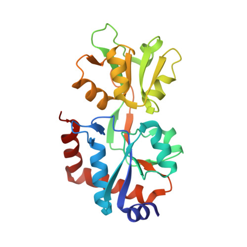
The non-swapped monomeric structure of the arginine-binding protein from Thermotoga maritima.
Smaldone, G., Ruggiero, A., Balasco, N., Abuhammad, A., Autiero, I., Caruso, D., Esposito, D., Ferraro, G., Gelardi, E.L.M., Moreira, M., Quareshy, M., Romano, M., Saaret, A., Selvam, I., Squeglia, F., Troisi, R., Kroon-Batenburg, L.M.J., Esposito, L., Berisio, R., Vitagliano, L.(2019) Acta Crystallogr F Struct Biol Commun 75: 707-713
- PubMed: 31702584 Search on PubMedSearch on PubMed Central
- DOI: https://doi.org/10.1107/S2053230X1901464X
- Primary Citation Related Structures:
6SVF - PubMed Abstract:
Domain swapping is a widespread oligomerization process that is observed in a large variety of protein families. In the large superfamily of substrate-binding proteins, non-monomeric members have rarely been reported. The arginine-binding protein from Thermotoga maritima (TmArgBP), a protein endowed with a number of unusual properties, presents a domain-swapped structure in its dimeric native state in which the two polypeptide chains mutually exchange their C-terminal helices. It has previously been shown that mutations in the region connecting the last two helices of the TmArgBP structure lead to the formation of a variety of oligomeric states (monomers, dimers, trimers and larger aggregates). With the aim of defining the structural determinants of domain swapping in TmArgBP, the monomeric form of the P235GK mutant has been structurally characterized. Analysis of this arginine-bound structure indicates that it consists of a closed monomer with its C-terminal helix folded against the rest of the protein, as typically observed for substrate-binding proteins. Notably, the two terminal helices are joined by a single nonhelical residue (Gly235). Collectively, the present findings indicate that extending the hinge region and conferring it with more conformational freedom makes the formation of a closed TmArgBP monomer possible. On the other hand, the short connection between the helices may explain the tendency of the protein to also adopt alternative oligomeric states (dimers, trimers and larger aggregates). The data reported here highlight the importance of evolutionary control to avoid the uncontrolled formation of heterogeneous and potentially harmful oligomeric species through domain swapping.
- AIC School Crystallographic Information Fiesta 2019, Naples, Italy.
Organizational Affiliation: