6SU3

Crystal structure of the 48C12 heliorhodopsin in the violet form at pH 8.8


Experimental Data Snapshot

  • Method: X-RAY DIFFRACTION
  • Resolution: 1.50 Å
  • R-Value Free: 0.199 
  • R-Value Work: 0.153 
  • R-Value Observed: 0.155 

wwPDB Validation   3D Report Full Report


Ligand Structure Quality Assessment 


This is version 2.1 of the entry. See complete history


Literature

High-resolution structural insights into the heliorhodopsin family.

Kovalev, K.Volkov, D.Astashkin, R.Alekseev, A.Gushchin, I.Haro-Moreno, J.M.Chizhov, I.Siletsky, S.Mamedov, M.Rogachev, A.Balandin, T.Borshchevskiy, V.Popov, A.Bourenkov, G.Bamberg, E.Rodriguez-Valera, F.Buldt, G.Gordeliy, V.

(2020) Proc Natl Acad Sci U S A 117: 4131-4141

  • DOI: https://doi.org/10.1073/pnas.1915888117
  • Primary Citation of Related Structures:  
    6SU3, 6SU4

  • PubMed Abstract: 

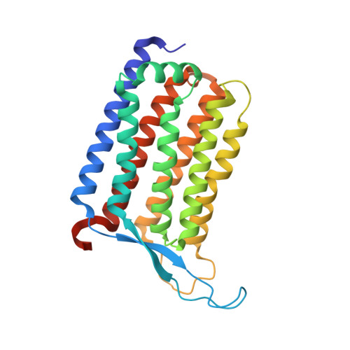
    Rhodopsins are the most abundant light-harvesting proteins. A new family of rhodopsins, heliorhodopsins (HeRs), has recently been discovered. Unlike in the known rhodopsins, in HeRs the N termini face the cytoplasm. The function of HeRs remains unknown. We present the structures of the bacterial HeR-48C12 in two states at the resolution of 1.5 Å, which highlight its remarkable difference from all known rhodopsins. The interior of HeR's extracellular part is completely hydrophobic, while the cytoplasmic part comprises a cavity (Schiff base cavity [SBC]) surrounded by charged amino acids and containing a cluster of water molecules, presumably being a primary proton acceptor from the Schiff base. At acidic pH, a planar triangular molecule (acetate) is present in the SBC. Structure-based bioinformatic analysis identified 10 subfamilies of HeRs, suggesting their diverse biological functions. The structures and available data suggest an enzymatic activity of HeR-48C12 subfamily and their possible involvement in fundamental redox biological processes.


  • Organizational Affiliation

    Institut de Biologie Structurale J.-P. Ebel, Université Grenoble Alpes-Commission for Atomic Energy (CEA)-CNRS, 38000 Grenoble, France.


Macromolecules
Find similar proteins by:  (by identity cutoff)  |  3D Structure
Entity ID: 1
MoleculeChains Sequence LengthOrganismDetailsImage
48C12 heliorhodopsinA [auth X],
B [auth A]
264Actinomycetes bacteriumMutation(s): 0 
Membrane Entity: Yes 
UniProt
Find proteins for A0A2R4S913 (uncultured Actinomycetes bacterium)
Explore A0A2R4S913 
Go to UniProtKB:  A0A2R4S913
Entity Groups  
Sequence Clusters30% Identity50% Identity70% Identity90% Identity95% Identity100% Identity
UniProt GroupA0A2R4S913
Sequence Annotations
Expand
  • Reference Sequence
Small Molecules
Ligands 6 Unique
IDChains Name / Formula / InChI Key2D Diagram3D Interactions
OLC
Query on OLC

Download Ideal Coordinates CCD File 
D [auth X],
E [auth X],
W [auth A]
(2R)-2,3-dihydroxypropyl (9Z)-octadec-9-enoate
C21 H40 O4
RZRNAYUHWVFMIP-GDCKJWNLSA-N
RET
Query on RET

Download Ideal Coordinates CCD File 
KA [auth A],
S [auth X]
RETINAL
C20 H28 O
NCYCYZXNIZJOKI-OVSJKPMPSA-N
LFA
Query on LFA

Download Ideal Coordinates CCD File 
AA [auth A]
BA [auth A]
C [auth X]
CA [auth A]
DA [auth A]
AA [auth A],
BA [auth A],
C [auth X],
CA [auth A],
DA [auth A],
EA [auth A],
FA [auth A],
G [auth X],
GA [auth A],
H [auth X],
HA [auth A],
I [auth X],
IA [auth A],
J [auth X],
JA [auth A],
K [auth X],
L [auth X],
M [auth X],
N [auth X],
O [auth X],
P [auth X],
Q [auth X],
R [auth X],
X [auth A],
Y [auth A],
Z [auth A]
EICOSANE
C20 H42
CBFCDTFDPHXCNY-UHFFFAOYSA-N
OLA
Query on OLA

Download Ideal Coordinates CCD File 
U [auth A],
V [auth A]
OLEIC ACID
C18 H34 O2
ZQPPMHVWECSIRJ-KTKRTIGZSA-N
SO4
Query on SO4

Download Ideal Coordinates CCD File 
F [auth X]SULFATE ION
O4 S
QAOWNCQODCNURD-UHFFFAOYSA-L
GOL
Query on GOL

Download Ideal Coordinates CCD File 
T [auth A]GLYCEROL
C3 H8 O3
PEDCQBHIVMGVHV-UHFFFAOYSA-N
Experimental Data & Validation

Experimental Data

  • Method: X-RAY DIFFRACTION
  • Resolution: 1.50 Å
  • R-Value Free: 0.199 
  • R-Value Work: 0.153 
  • R-Value Observed: 0.155 
  • Space Group: P 1 21 1
Unit Cell:
Length ( Å )Angle ( ˚ )
a = 56.115α = 90
b = 59.748β = 92.3
c = 94.318γ = 90
Software Package:
Software NamePurpose
REFMACrefinement
Aimlessdata scaling
PDB_EXTRACTdata extraction
XDSdata reduction
MOLREPphasing

Structure Validation

View Full Validation Report


Ligand Structure Quality Assessment 


Entry History & Funding Information

Deposition Data


Funding OrganizationLocationGrant Number
French National Research AgencyFranceANR-15-CE11-0029-02/FA 301/11-1
French National Research AgencyFranceANR-14-CE09-0028
French Infrastructure for Integrated Structural BiologyFranceANR-10-INSB-05-02
Grenoble Alliance for Integrated Structural Cell BiologyFranceANR-10-LABX-49-01
Russian Foundation for Basic ResearchRussian Federation19-52-15017

Revision History  (Full details and data files)

  • Version 1.0: 2019-12-11
    Type: Initial release
  • Version 1.1: 2020-02-19
    Changes: Database references
  • Version 1.2: 2020-03-04
    Changes: Database references
  • Version 2.0: 2020-12-02
    Changes: Advisory, Atomic model, Derived calculations, Refinement description
  • Version 2.1: 2024-01-24
    Changes: Data collection, Database references, Refinement description