6SQU

Crystal structure of human SHIP2 catalytic domain in complex with 1,2,4 Dimer


Experimental Data Snapshot

  • Method: X-RAY DIFFRACTION
  • Resolution: 2.27 Å
  • R-Value Free: 0.254 
  • R-Value Work: 0.201 
  • R-Value Observed: 0.203 

wwPDB Validation   3D Report Full Report


This is version 1.2 of the entry. See complete history


Literature

Allosteric Site on SHIP2 Identified Through Fluorescent Ligand Screening and Crystallography: A Potential New Target for Intervention.

Whitfield, H.Hemmings, A.M.Mills, S.J.Baker, K.White, G.Rushworth, S.Riley, A.M.Potter, B.V.L.Brearley, C.A.

(2021) J Med Chem 64: 3813-3826

  • DOI: https://doi.org/10.1021/acs.jmedchem.0c01944
  • Primary Citation of Related Structures:  
    6SQU, 6SRR

  • PubMed Abstract: 

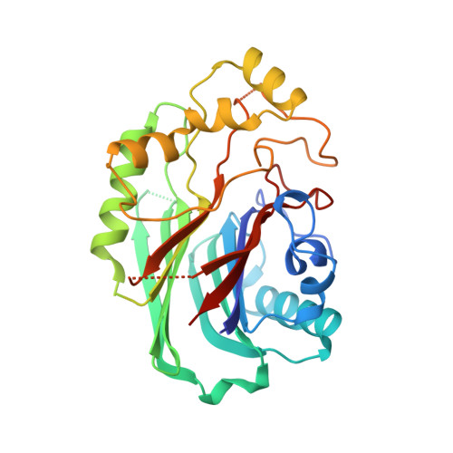
    Src homology 2 domain-containing inositol phosphate phosphatase 2 (SHIP2) is one of the 10 human inositol phosphate 5-phosphatases. One of its physiological functions is dephosphorylation of phosphatidylinositol 3,4,5-trisphosphate, PtdIns(3,4,5)P 3 . It is therefore a therapeutic target for pathophysiologies dependent on PtdIns(3,4,5)P 3 and PtdIns(3,4)P 2 . Therapeutic interventions are limited by the dearth of crystallographic data describing ligand/inhibitor binding. An active site-directed fluorescent probe facilitated screening of compound libraries for SHIP2 ligands. With two additional orthogonal assays, several ligands including galloflavin were identified as low micromolar Ki inhibitors. One ligand, an oxo-linked ethylene-bridged dimer of benzene 1,2,4-trisphosphate, was shown to be an uncompetitive inhibitor that binds to a regulatory site on the catalytic domain. We posit that binding of ligands to this site restrains L4 loop motions that are key to interdomain communications that accompany high catalytic activity with phosphoinositide substrate. This site may, therefore, be a future druggable target for medicinal chemistry.


  • Organizational Affiliation

    School of Biological Sciences, University of East Anglia, Norwich Research Park, Norwich NR4 7TJ, U.K.


Macromolecules
Find similar proteins by:  (by identity cutoff)  |  3D Structure
Entity ID: 1
MoleculeChains Sequence LengthOrganismDetailsImage
Phosphatidylinositol 3,4,5-trisphosphate 5-phosphatase 2
A, B
316Homo sapiensMutation(s): 0 
Gene Names: INPPL1SHIP2
EC: 3.1.3.86
UniProt & NIH Common Fund Data Resources
Find proteins for O15357 (Homo sapiens)
Explore O15357 
Go to UniProtKB:  O15357
PHAROS:  O15357
GTEx:  ENSG00000165458 
Entity Groups  
Sequence Clusters30% Identity50% Identity70% Identity90% Identity95% Identity100% Identity
UniProt GroupO15357
Sequence Annotations
Expand
  • Reference Sequence
Small Molecules
Ligands 1 Unique
IDChains Name / Formula / InChI Key2D Diagram3D Interactions
D7I (Subject of Investigation/LOI)
Query on D7I

Download Ideal Coordinates CCD File 
C [auth B]5,5'-(ethane-1,2-diylbis(oxy))bis(benzene-5,4,2,1,-tetrayl)hexakisphosphate
C14 H20 O26 P6
SPZGMBGXCYAMCB-UHFFFAOYSA-N
Experimental Data & Validation

Experimental Data

  • Method: X-RAY DIFFRACTION
  • Resolution: 2.27 Å
  • R-Value Free: 0.254 
  • R-Value Work: 0.201 
  • R-Value Observed: 0.203 
  • Space Group: P 1 21 1
Unit Cell:
Length ( Å )Angle ( ˚ )
a = 44.41α = 90
b = 60.19β = 92.75
c = 113.96γ = 90
Software Package:
Software NamePurpose
PHENIXrefinement
xia2data reduction
xia2data scaling
PHASERphasing

Structure Validation

View Full Validation Report


Entry History 

Deposition Data

Revision History  (Full details and data files)

  • Version 1.0: 2021-01-13
    Type: Initial release
  • Version 1.1: 2021-07-28
    Changes: Database references
  • Version 1.2: 2024-01-24
    Changes: Data collection, Database references, Refinement description