


Crystal structure of mouse PRMT6 in complex with inhibitors
Bonnefond, L., Cavarelli, J.To be published.
Experimental Data Snapshot
Starting Model: experimental
View more details
wwPDB Validation 3D Report Full Report

Entity ID: 1 | |||||
|---|---|---|---|---|---|
| Molecule | Chains | Sequence Length | Organism | Details | Image |
| Protein arginine N-methyltransferase 6 | 380 | Mus musculus | Mutation(s): 1 Gene Names: Prmt6, Hrmt1l6 EC: 2.1.1.319 | 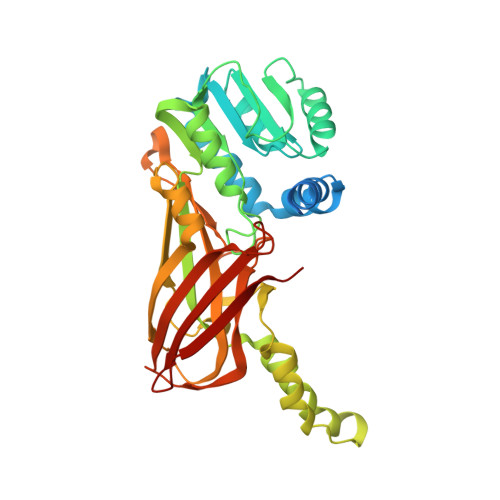 | |
UniProt & NIH Common Fund Data Resources | |||||
IMPC: MGI:2139971 | |||||
Entity Groups | |||||
| Sequence Clusters | 30% Identity50% Identity70% Identity90% Identity95% Identity100% Identity | ||||
| UniProt Group | Q6NZB1 | ||||
Sequence AnnotationsExpand | |||||
Reference Sequence | |||||
Entity ID: 2 | |||||
|---|---|---|---|---|---|
| Molecule | Chains | Sequence Length | Organism | Details | Image |
| H4-7 | C [auth D], D [auth E] | 7 | Homo sapiens | Mutation(s): 0 EC: 2.1.1.43 | 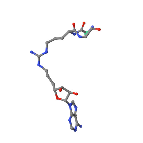 |
| Length ( Å ) | Angle ( ˚ ) |
|---|---|
| a = 118.632 | α = 90 |
| b = 142.829 | β = 90 |
| c = 41.701 | γ = 90 |
| Software Name | Purpose |
|---|---|
| XDS | data reduction |
| Aimless | data scaling |
| PHASER | phasing |
| PHENIX | refinement |
| PDB_EXTRACT | data extraction |


