6SQ7

Crystal structure of M. tuberculosis InhA in complex with NAD+ and 2-(4-chloro-3-nitrobenzoyl)benzoic acid


Experimental Data Snapshot

  • Method: X-RAY DIFFRACTION
  • Resolution: 1.76 Å
  • R-Value Free: 0.164 
  • R-Value Work: 0.156 
  • R-Value Observed: 0.157 

wwPDB Validation   3D Report Full Report


Ligand Structure Quality Assessment 


This is version 1.3 of the entry. See complete history


Literature

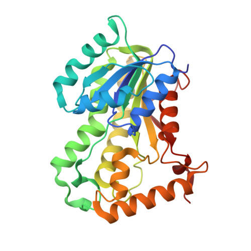
Fragment-Based Design ofMycobacterium tuberculosisInhA Inhibitors.

Sabbah, M.Mendes, V.Vistal, R.G.Dias, D.M.G.Zahorszka, M.Mikusova, K.Kordulakova, J.Coyne, A.G.Blundell, T.L.Abell, C.

(2020) J Med Chem 63: 4749-4761

  • DOI: https://doi.org/10.1021/acs.jmedchem.0c00007
  • Primary Citation of Related Structures:  
    6SQ5, 6SQ7, 6SQ9, 6SQB, 6SQD, 6SQL

  • PubMed Abstract: 

    Tuberculosis (TB) remains a leading cause of mortality among infectious diseases worldwide. InhA has been the focus of numerous drug discovery efforts as this is the target of the first line pro-drug isoniazid. However, with resistance to this drug becoming more common, the aim has been to find new clinical candidates that directly inhibit this enzyme and that do not require activation by the catalase peroxidase KatG, thus circumventing the majority of the resistance mechanisms. In this work, the screening and validation of a fragment library are described, and the development of the fragment hits using a fragment growing strategy was employed, which led to the development of InhA inhibitors with affinities of up to 250 nM.


  • Organizational Affiliation

    Department of Chemistry, University of Cambridge, Lensfield Road, Cambridge CB2 1EW, United Kingdom.


Macromolecules
Find similar proteins by:  (by identity cutoff)  |  3D Structure
Entity ID: 1
MoleculeChains Sequence LengthOrganismDetailsImage
Enoyl-[acyl-carrier-protein] reductase [NADH]270Mycobacterium tuberculosis H37RvMutation(s): 0 
Gene Names: inhARv1484MTCY277.05
EC: 1.3.1.9
UniProt
Find proteins for P9WGR1 (Mycobacterium tuberculosis (strain ATCC 25618 / H37Rv))
Explore P9WGR1 
Go to UniProtKB:  P9WGR1
Entity Groups  
Sequence Clusters30% Identity50% Identity70% Identity90% Identity95% Identity100% Identity
UniProt GroupP9WGR1
Sequence Annotations
Expand
  • Reference Sequence
Small Molecules
Ligands 2 Unique
IDChains Name / Formula / InChI Key2D Diagram3D Interactions
NAD
Query on NAD

Download Ideal Coordinates CCD File 
B [auth A]NICOTINAMIDE-ADENINE-DINUCLEOTIDE
C21 H27 N7 O14 P2
BAWFJGJZGIEFAR-NNYOXOHSSA-N
9FN (Subject of Investigation/LOI)
Query on 9FN

Download Ideal Coordinates CCD File 
C [auth A]2-(4-chloranyl-3-nitro-phenyl)carbonylbenzoic acid
C14 H8 Cl N O5
RITAQDHCJBLSSL-UHFFFAOYSA-N
Experimental Data & Validation

Experimental Data

  • Method: X-RAY DIFFRACTION
  • Resolution: 1.76 Å
  • R-Value Free: 0.164 
  • R-Value Work: 0.156 
  • R-Value Observed: 0.157 
  • Space Group: P 62 2 2
Unit Cell:
Length ( Å )Angle ( ˚ )
a = 97.524α = 90
b = 97.524β = 90
c = 140.181γ = 120
Software Package:
Software NamePurpose
PHENIXrefinement
Aimlessdata scaling
PHASERphasing
PDB_EXTRACTdata extraction
XDSdata reduction

Structure Validation

View Full Validation Report


Ligand Structure Quality Assessment 


Entry History & Funding Information

Deposition Data


Funding OrganizationLocationGrant Number
European Communitys Seventh Framework ProgrammeUnited Kingdom260872
Bill & Melinda Gates FoundationUnited StatesOPP1158806

Revision History  (Full details and data files)

  • Version 1.0: 2020-04-22
    Type: Initial release
  • Version 1.1: 2020-04-29
    Changes: Database references
  • Version 1.2: 2020-05-27
    Changes: Database references
  • Version 1.3: 2024-01-24
    Changes: Data collection, Database references, Refinement description