


Around she goes: the structure of mouse Interleukin-12 p80
Bloch, Y., Savvides, S.N.To be published.
Experimental Data Snapshot
Starting Model: experimental
View more details
wwPDB Validation 3D Report Full Report

A newer entry is available that reflects an alternative modeling of the original data: 7PUR
Entity ID: 1 | |||||
|---|---|---|---|---|---|
| Molecule | Chains | Sequence Length | Organism | Details | Image |
| Interleukin-12 subunit beta | 344 | Mus musculus | Mutation(s): 0 Gene Names: Il12b | 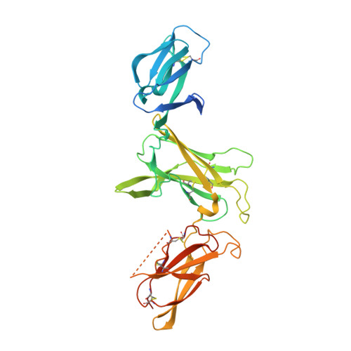 | |
UniProt & NIH Common Fund Data Resources | |||||
IMPC: MGI:96540 | |||||
Entity Groups | |||||
| Sequence Clusters | 30% Identity50% Identity70% Identity90% Identity95% Identity100% Identity | ||||
| UniProt Group | P43432 | ||||
Glycosylation | |||||
| Glycosylation Sites: 1 | Go to GlyGen: P43432-1 | ||||
Sequence AnnotationsExpand | |||||
Reference Sequence | |||||
Entity ID: 2 | |||||
|---|---|---|---|---|---|
| Molecule | Chains | Length | 2D Diagram | Glycosylation | D Interactions |
| alpha-D-mannopyranose-(1-3)-alpha-D-mannopyranose-(1-6)-[alpha-D-mannopyranose-(1-3)]beta-D-mannopyranose-(1-4)-2-acetamido-2-deoxy-beta-D-glucopyranose-(1-4)-2-acetamido-2-deoxy-beta-D-glucopyranose | C, D | 6 |  | N-Glycosylation | |
Glycosylation Resources | |||||
GlyTouCan: G09724ZC GlyCosmos: G09724ZC GlyGen: G09724ZC | |||||
| Ligands 1 Unique | |||||
|---|---|---|---|---|---|
| ID | Chains | Name / Formula / InChI Key | 2D Diagram | 3D Interactions | |
| CL Download:Ideal Coordinates CCD File | E [auth B] | CHLORIDE ION Cl VEXZGXHMUGYJMC-UHFFFAOYSA-M |  | ||
| Length ( Å ) | Angle ( ˚ ) |
|---|---|
| a = 53.8 | α = 90 |
| b = 176.14 | β = 108.963 |
| c = 54.37 | γ = 90 |
| Software Name | Purpose |
|---|---|
| BUSTER | refinement |
| PHENIX | refinement |
| XDS | data reduction |
| XDS | data scaling |
| PHASER | phasing |
| Funding Organization | Location | Grant Number |
|---|---|---|
| Research Foundation - Flanders | Belgium | 12S0519N |
| Research Foundation - Flanders | Belgium | G0B4918N |
| Research Foundation - Flanders | Belgium | G0E1516N |


