6SL9 | pdb_00006sl9

High resolution apo structure of isomerase PaaG


Experimental Data Snapshot

  • Method: X-RAY DIFFRACTION
  • Resolution: 1.27 Å
  • R-Value Free: 
    0.179 (Depositor), 0.178 (DCC) 
  • R-Value Work: 
    0.146 (Depositor) 

Starting Model: experimental
View more details

wwPDB Validation 3D Report Full Report

Validation slider image for 6SL9

This is version 1.2 of the entry. See complete history

Literature

Structural and Mechanistic Basis of an Oxepin-CoA Forming Isomerase in Bacterial Primary and Secondary Metabolism.

Spieker, M.Saleem-Batcha, R.Teufel, R.

(2019) ACS Chem Biol 14: 2876-2886

  • DOI: https://doi.org/10.1021/acschembio.9b00742
  • Primary Citation Related Structures: 
    6SL9, 6SLA, 6SLB

  • PubMed Abstract: 

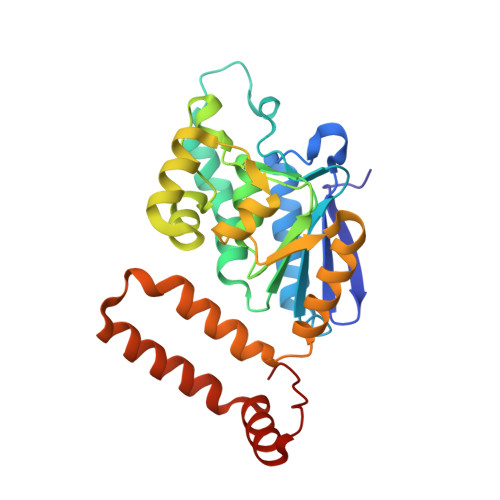
    Numerous aromatic compounds are aerobically degraded in bacteria via the central intermediate phenylacetic acid (paa). In one of the key steps of this widespread catabolic pathway, 1,2-epoxyphenylacetyl-CoA is converted by PaaG into the heterocyclic oxepin-CoA. PaaG thereby elegantly generates an α,β-unsaturated CoA ester that is predisposed to undergo β-oxidation subsequent to hydrolytic ring-cleavage. Moreover, oxepin-CoA serves as a precursor for secondary metabolites (e.g., tropodithietic acid) that act as antibiotics and quorum-sensing signals. Here we verify that PaaG adopts a second role in aromatic catabolism by converting cis -3,4-didehydroadipoyl-CoA into trans -2,3-didehydroadipoyl-CoA and corroborate a Δ 3 2 -enoyl-CoA isomerase-like proton shuttling mechanism for both distinct substrates. Biochemical and structural investigations of PaaG reveal active site adaptations to the structurally different substrates and provide detailed insight into catalysis and control of stereospecificity. This work elucidates the mechanism of action of unusual isomerase PaaG and sheds new light on the ubiquitous enoyl-CoA isomerases of the crotonase superfamily.


  • Organizational Affiliation
    • ZBSA, Center for Biological Systems Analysis , University of Freiburg , 79104 Freiburg , Germany.

Macromolecule Content 

  • Total Structure Weight: 28.13 kDa 
  • Atom Count: 2,180 
  • Modeled Residue Count: 258 
  • Deposited Residue Count: 258 
  • Unique protein chains: 1

Macromolecules

Find similar proteins by:|  3D Structure
Entity ID: 1
MoleculeChains  Sequence LengthOrganismDetailsImage
Enoyl-CoA hydratase/carnithine racemaseA [auth AAA]258Thermus thermophilus JL-18Mutation(s): 0 
Gene Names: TtJL18_0099
UniProt
Find proteins for H9ZNW0 (Thermus thermophilus JL-18)
Explore H9ZNW0 
Go to UniProtKB:  H9ZNW0
Entity Groups
Sequence Clusters30% Identity50% Identity70% Identity90% Identity95% Identity100% Identity
UniProt GroupH9ZNW0
Sequence Annotations
Expand
Reference Sequence

Experimental Data & Validation

Experimental Data

  • Method: X-RAY DIFFRACTION
  • Resolution: 1.27 Å
  • R-Value Free:  0.179 (Depositor), 0.178 (DCC) 
  • R-Value Work:  0.146 (Depositor) 
Space Group: P 21 3
Unit Cell:
Length ( Å )Angle ( ˚ )
a = 88.497α = 90
b = 88.497β = 90
c = 88.497γ = 90
Software Package:
Software NamePurpose
REFMACrefinement
XDSdata reduction
SCALAdata scaling
PHASERphasing

Structure Validation

View Full Validation Report


Entry History 

& Funding Information

Deposition Data


Funding OrganizationLocationGrant Number
German Research FoundationGermanyTE 931/2-1

Revision History  (Full details and data files)

  • Version 1.0: 2019-12-11
    Type: Initial release
  • Version 1.1: 2020-01-01
    Changes: Database references
  • Version 1.2: 2024-01-24
    Changes: Data collection, Database references, Derived calculations, Refinement description