Thermodynamic and Evolutionary Coupling between the Native and Amyloid State of Globular Proteins.
Langenberg, T., Gallardo, R., van der Kant, R., Louros, N., Michiels, E., Duran-Romana, R., Houben, B., Cassio, R., Wilkinson, H., Garcia, T., Ulens, C., Van Durme, J., Rousseau, F., Schymkowitz, J.(2020) Cell Rep 31: 107512-107512
- PubMed: 32294448 Search on PubMedSearch on PubMed Central
- DOI: https://doi.org/10.1016/j.celrep.2020.03.076
- Primary Citation Related Structures:
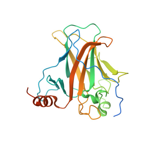
6SL6 - PubMed Abstract:
The amyloid-like aggregation propensity present in most globular proteins is generally considered to be a secondary side effect resulting from the requirements of protein stability. Here, we demonstrate, however, that mutations in the globular and amyloid state are thermodynamically correlated rather than simply associated. In addition, we show that the standard genetic code couples this structural correlation into a tight evolutionary relationship. We illustrate the extent of this evolutionary entanglement of amyloid propensity and globular protein stability. Suppressing a 600-Ma-conserved amyloidogenic segment in the p53 core domain fold is structurally feasible but requires 7-bp substitutions to concomitantly introduce two aggregation-suppressing and three stabilizing amino acid mutations. We speculate that, rather than being a corollary of protein evolution, it is equally plausible that positive selection for amyloid structure could have been a driver for the emergence of globular protein structure.
- Switch Laboratory, VIB Center for Brain and Disease Research, Herestraat 49, 3000 Leuven, Belgium; Switch Laboratory, Department of Cellular and Molecular Medicine, KU Leuven, Herestraat 49, 3000 Leuven, Belgium.
Organizational Affiliation: