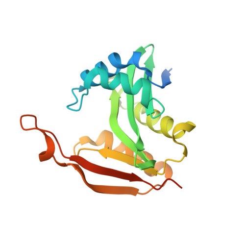
The architecture of the diaminobutyrate acetyltransferase active site provides mechanistic insight into the biosynthesis of the chemical chaperone ectoine.
Richter, A.A., Kobus, S., Czech, L., Hoeppner, A., Zarzycki, J., Erb, T.J., Lauterbach, L., Dickschat, J.S., Bremer, E., Smits, S.H.J.(2020) J Biological Chem 295: 2822-2838
- PubMed: 31969391 Search on PubMedSearch on PubMed Central
- DOI: https://doi.org/10.1074/jbc.RA119.011277
- Primary Citation Related Structures:
6SJY, 6SK1, 6SL8, 6SLK, 6SLL - PubMed Abstract:
Ectoine is a solute compatible with the physiologies of both prokaryotic and eukaryotic cells and is widely synthesized by bacteria as an osmotic stress protectant. Because it preserves functional attributes of proteins and macromolecular complexes, it is considered a chemical chaperone and has found numerous practical applications. However, the mechanism of its biosynthesis is incompletely understood. The second step in ectoine biosynthesis is catalyzed by l-2,4-diaminobutyrate acetyltransferase (EctA; EC 2.3.1.178), which transfers the acetyl group from acetyl-CoA to EctB-formed l-2,4-diaminobutyrate (DAB), yielding N -γ-acetyl-l-2,4-diaminobutyrate ( N -γ-ADABA), the substrate of ectoine synthase (EctC). Here, we report the biochemical and structural characterization of the EctA enzyme from the thermotolerant bacterium Paenibacillus lautus ( Pl ). We found that ( Pl )EctA forms a homodimer whose enzyme activity is highly regiospecific by producing N -γ-ADABA but not the ectoine catabolic intermediate N -α-acetyl-l-2,4-diaminobutyric acid. High-resolution crystal structures of ( Pl )EctA (at 1.2-2.2 Å resolution) (i) for its apo-form, (ii) in complex with CoA, (iii) in complex with DAB, (iv) in complex with both CoA and DAB, and (v) in the presence of the product N -γ-ADABA were obtained. To pinpoint residues involved in DAB binding, we probed the structure-function relationship of ( Pl )EctA by site-directed mutagenesis. Phylogenomics shows that EctA-type proteins from both Bacteria and Archaea are evolutionarily highly conserved, including catalytically important residues. Collectively, our biochemical and structural findings yielded detailed insights into the catalytic core of the EctA enzyme that laid the foundation for unraveling its reaction mechanism.
- Department of Biology, Laboratory for Microbiology, Philipps-University Marburg, D-35043 Marburg, Germany; SYNMIKRO Research Center, Philipps-University Marburg, D-35043 Marburg, Germany.
Organizational Affiliation: