Download Mendeley
The type 2 diabetes gene product STARD10 is a phosphoinositide-binding protein that controls insulin secretory granule biogenesis.
Carrat, G.R., Haythorne, E., Tomas, A., Haataja, L., Muller, A., Arvan, P., Piunti, A., Cheng, K., Huang, M., Pullen, T.J., Georgiadou, E., Stylianides, T., Amirruddin, N.S., Salem, V., Distaso, W., Cakebread, A., Heesom, K.J., Lewis, P.A., Hodson, D.J., Briant, L.J., Fung, A.C.H., Sessions, R.B., Alpy, F., Kong, A.P.S., Benke, P.I., Torta, F., Teo, A.K.K., Leclerc, I., Solimena, M., Wigley, D.B., Rutter, G.A.(2020) Mol Metab 40: 101015-101015
- PubMed: 32416313 Search on PubMedSearch on PubMed Central
- DOI: https://doi.org/10.1016/j.molmet.2020.101015
- Primary Citation Related Structures:
6SER - PubMed Abstract:
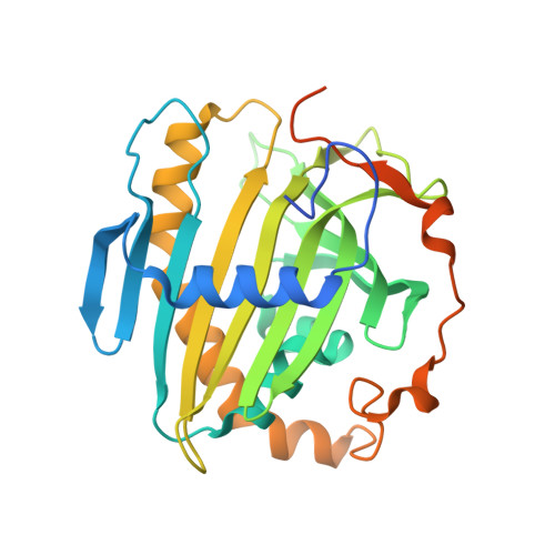
Risk alleles for type 2 diabetes at the STARD10 locus are associated with lowered STARD10 expression in the β-cell, impaired glucose-induced insulin secretion, and decreased circulating proinsulin:insulin ratios. Although likely to serve as a mediator of intracellular lipid transfer, the identity of the transported lipids and thus the pathways through which STARD10 regulates β-cell function are not understood. The aim of this study was to identify the lipids transported and affected by STARD10 in the β-cell and the role of the protein in controlling proinsulin processing and insulin granule biogenesis and maturation. We used isolated islets from mice deleted selectively in the β-cell for Stard10 (βStard10KO) and performed electron microscopy, pulse-chase, RNA sequencing, and lipidomic analyses. Proteomic analysis of STARD10 binding partners was executed in the INS1 (832/13) cell line. X-ray crystallography followed by molecular docking and lipid overlay assay was performed on purified STARD10 protein. βStard10KO islets had a sharply altered dense core granule appearance, with a dramatic increase in the number of "rod-like" dense cores. Correspondingly, basal secretion of proinsulin was increased versus wild-type islets. The solution of the crystal structure of STARD10 to 2.3 Å resolution revealed a binding pocket capable of accommodating polyphosphoinositides, and STARD10 was shown to bind to inositides phosphorylated at the 3' position. Lipidomic analysis of βStard10KO islets demonstrated changes in phosphatidylinositol levels, and the inositol lipid kinase PIP4K2C was identified as a STARD10 binding partner. Also consistent with roles for STARD10 in phosphoinositide signalling, the phosphoinositide-binding proteins Pirt and Synaptotagmin 1 were amongst the differentially expressed genes in βStard10KO islets. Our data indicate that STARD10 binds to, and may transport, phosphatidylinositides, influencing membrane lipid composition, insulin granule biosynthesis, and insulin processing.
- Section of Cell Biology and Functional Genomics, Imperial College London, du Cane Road, London, W12 0NN, UK.
Organizational Affiliation: