


Crystallographic Fragment Screening of the parasitic PEX14
Hassaan, E., Heine, A., Klebe, G.To be published.
Experimental Data Snapshot
wwPDB Validation 3D Report Full Report

Entity ID: 1 | |||||
|---|---|---|---|---|---|
| Molecule | Chains | Sequence Length | Organism | Details | Image |
| Peroxin 14 | 66 | Trypanosoma brucei brucei | Mutation(s): 0 Gene Names: PEX14 | 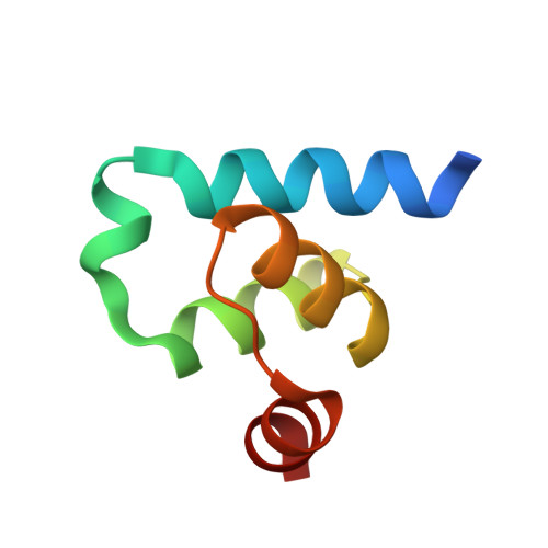 | |
UniProt | |||||
Entity Groups | |||||
| Sequence Clusters | 30% Identity50% Identity70% Identity90% Identity95% Identity100% Identity | ||||
| UniProt Group | Q38CL4 | ||||
Sequence AnnotationsExpand | |||||
Reference Sequence | |||||
| Ligands 1 Unique | |||||
|---|---|---|---|---|---|
| ID | Chains | Name / Formula / InChI Key | 2D Diagram | 3D Interactions | |
| PEG Download:Ideal Coordinates CCD File | E [auth C], F [auth D] | DI(HYDROXYETHYL)ETHER C4 H10 O3 MTHSVFCYNBDYFN-UHFFFAOYSA-N |  | ||
| Length ( Å ) | Angle ( ˚ ) |
|---|---|
| a = 38.925 | α = 90 |
| b = 81.268 | β = 92.75 |
| c = 39.287 | γ = 90 |
| Software Name | Purpose |
|---|---|
| PHENIX | refinement |
| PHASER | phasing |
| Coot | model building |
| XDS | data scaling |
| XDS | data reduction |
| Funding Organization | Location | Grant Number |
|---|---|---|
| European Commission | Germany | -- |


