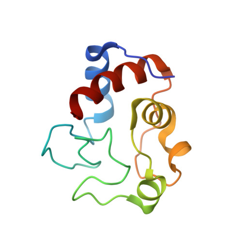
6S8Y

Crystal structure of cytochrome c in complex with a sulfonated quinoline-derived foldamer


Experimental Data Snapshot

  • Method: X-RAY DIFFRACTION
  • Resolution: 2.09 Å
  • R-Value Free: 0.267 
  • R-Value Work: 0.225 
  • R-Value Observed: 0.227 

wwPDB Validation   3D Report Full Report


Ligand Structure Quality Assessment 


This is version 1.0 of the entry. See complete history


Literature

Crystal structure of a protein-aromatic foldamer composite: macromolecular chiral resolution.

Alex, J.M.Corvaglia, V.Hu, X.Engilberge, S.Huc, I.Crowley, P.B.

(2019) Chem Commun (Camb) 55: 11087-11090

  • DOI: https://doi.org/10.1039/c9cc05330a
  • Primary Citation of Related Structures:  
    6S8Y

  • PubMed Abstract: 
  • Co-crystallization of a 2 kDa tether-free sulfonated foldamer and the 13 kDa lysine-rich cytochrome c yielded a remarkable biohybrid assembly with chiral resolution of the foldamer helix handedness. In the crystal a ∼5 nm foldamer stack was surrounded by eight molecules of protein ...

    Co-crystallization of a 2 kDa tether-free sulfonated foldamer and the 13 kDa lysine-rich cytochrome c yielded a remarkable biohybrid assembly with chiral resolution of the foldamer helix handedness. In the crystal a ∼5 nm foldamer stack was surrounded by eight molecules of protein. NMR and CD experiments suggest interesting differences in the solution state recognition processes.


    Organizational Affiliation

    School of Chemistry, National University of Ireland, University Road, Galway, H91 TK33, Ireland.


Macromolecules
Find similar proteins by:  (by identity cutoff)  |  3D Structure
Entity ID: 1
MoleculeChainsSequence LengthOrganismDetailsImage
Cytochrome c iso-1107Saccharomyces cerevisiaeMutation(s): 0 
Gene Names: CYC1YJR048WJ1653
UniProt
Find proteins for P00044 (Saccharomyces cerevisiae (strain ATCC 204508 / S288c))
Explore P00044 
Go to UniProtKB:  P00044
Entity Groups  
Sequence Clusters30% Identity50% Identity70% Identity90% Identity95% Identity100% Identity
UniProt GroupP00044
Protein Feature View
Expand
  • Reference Sequence
Small Molecules
Ligands 4 Unique
IDChainsName / Formula / InChI Key2D Diagram3D Interactions
L0T (Subject of Investigation/LOI)
Query on L0T

Download Ideal Coordinates CCD File 
G [auth A],
H [auth A]
8-acetamido-2-[[2-[[2-[[2-[[2-[[2-[[2-[(2-carboxy-4-sulfonato-quinolin-8-yl)carbamoyl]-4-sulfonato-quinolin-8-yl]carbamoyl]-4-sulfonato-quinolin-8-yl]carbamoyl]-4-sulfonato-quinolin-8-yl]carbamoyl]-4-sulfonato-quinolin-8-yl]carbamoyl]-4-sulfonato-quinolin-8-yl]carbamoyl]-4-sulfonato-quinolin-8-yl]carbamoyl]quinoline-4-sulfonate
C82 H44 N16 O34 S8
IHZXKZSCEKIUSH-UHFFFAOYSA-F
 Ligand Interaction
HEC
Query on HEC

Download Ideal Coordinates CCD File 
B [auth A]HEME C
C34 H34 Fe N4 O4
HXQIYSLZKNYNMH-LJNAALQVSA-N
 Ligand Interaction
ZN
Query on ZN

Download Ideal Coordinates CCD File 
C [auth A],
D [auth A],
I [auth A]
ZINC ION
Zn
PTFCDOFLOPIGGS-UHFFFAOYSA-N
 Ligand Interaction
ACT
Query on ACT

Download Ideal Coordinates CCD File 
E [auth A],
F [auth A]
ACETATE ION
C2 H3 O2
QTBSBXVTEAMEQO-UHFFFAOYSA-M
 Ligand Interaction
Experimental Data & Validation

Experimental Data

  • Method: X-RAY DIFFRACTION
  • Resolution: 2.09 Å
  • R-Value Free: 0.267 
  • R-Value Work: 0.225 
  • R-Value Observed: 0.227 
  • Space Group: P 32 2 1
Unit Cell:
Length ( Å )Angle ( ˚ )
a = 39.138α = 90
b = 39.138β = 90
c = 187.366γ = 120
Software Package:
Software NamePurpose
BUSTERrefinement
PDB_EXTRACTdata extraction
XDSdata reduction
Aimlessdata scaling
MOLREPphasing

Structure Validation

View Full Validation Report


Ligand Structure Quality Assessment 


Entry History & Funding Information

Deposition Data


Funding OrganizationLocationGrant Number
Science Foundation IrelandIreland13/CDA/2168

Revision History  (Full details and data files)

  • Version 1.0: 2019-09-25
    Type: Initial release