6S83

Crystal structure of methionine adenosyltransferase from Pyrococcus furiosus in complex with AMPPCP, SAM, and PCP


Experimental Data Snapshot

  • Method: X-RAY DIFFRACTION
  • Resolution: 2.34 Å
  • R-Value Free: 0.240 
  • R-Value Work: 0.207 
  • R-Value Observed: 0.209 

wwPDB Validation   3D Report Full Report


Ligand Structure Quality Assessment 


This is version 1.2 of the entry. See complete history


Literature

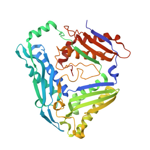
Structures of catalytic cycle intermediates of the Pyrococcus furiosus methionine adenosyltransferase demonstrate negative cooperativity in the archaeal orthologues.

Minici, C.Mosca, L.Ilisso, C.P.Cacciapuoti, G.Porcelli, M.Degano, M.

(2020) J Struct Biol 210: 107462-107462

  • DOI: https://doi.org/10.1016/j.jsb.2020.107462
  • Primary Citation of Related Structures:  
    6S81, 6S83

  • PubMed Abstract: 

    Methionine adenosyltransferases catalyse the biosynthesis of S-adenosylmethionine, the primary methyl group donor in biochemical reactions, through the condensation of methionine and ATP. Here, we report the structural analysis of the Pyrococcus furiosus methionine adenosyltransferase (PfMAT) captured in the unliganded, substrate- and product-bound states. The conformational changes taking place during the enzymatic catalytic cycle are allosterically propagated by amino acid residues conserved in the archaeal orthologues to induce an asymmetric dimer structure. The distinct occupancy of the active sites within a PfMAT dimer is consistent with a half-site reactivity that is mediated by a product-induced negative cooperativity. The structures of intermediate states of PfMAT reported here suggest a distinct molecular mechanism for S-adenosylmethionine synthesis in Archaea, likely consequence of the evolutionary pressure to achieve protein stability under extreme conditions.


  • Organizational Affiliation

    Biocrystallography Unit, Division of Immunology, Transplantation, and Infectious Diseases, IRCCS Scientific Institute San Raffaele, 20132 Milan, Italy.


Macromolecules
Find similar proteins by:  (by identity cutoff)  |  3D Structure
Entity ID: 1
MoleculeChains Sequence LengthOrganismDetailsImage
S-adenosylmethionine synthase
A, B, C, D, E
A, B, C, D, E, F, G, H
401Pyrococcus furiosus DSM 3638Mutation(s): 0 
Gene Names: matPF1866
EC: 2.5.1.6
UniProt
Find proteins for Q8TZW1 (Pyrococcus furiosus (strain ATCC 43587 / DSM 3638 / JCM 8422 / Vc1))
Explore Q8TZW1 
Go to UniProtKB:  Q8TZW1
Entity Groups  
Sequence Clusters30% Identity50% Identity70% Identity90% Identity95% Identity100% Identity
UniProt GroupQ8TZW1
Sequence Annotations
Expand
  • Reference Sequence
Small Molecules
Ligands 5 Unique
IDChains Name / Formula / InChI Key2D Diagram3D Interactions
ACP (Subject of Investigation/LOI)
Query on ACP

Download Ideal Coordinates CCD File 
IA [auth F],
P [auth B],
RA [auth H],
Y [auth D]
PHOSPHOMETHYLPHOSPHONIC ACID ADENYLATE ESTER
C11 H18 N5 O12 P3
UFZTZBNSLXELAL-IOSLPCCCSA-N
SAM (Subject of Investigation/LOI)
Query on SAM

Download Ideal Coordinates CCD File 
EA [auth E],
K [auth A]
S-ADENOSYLMETHIONINE
C15 H22 N6 O5 S
MEFKEPWMEQBLKI-FCKMPRQPSA-N
MDN (Subject of Investigation/LOI)
Query on MDN

Download Ideal Coordinates CCD File 
CA [auth E],
I [auth A],
OA [auth H],
T [auth C]
METHYLENEDIPHOSPHONIC ACID
C H6 O6 P2
MBKDYNNUVRNNRF-UHFFFAOYSA-N
PO4
Query on PO4

Download Ideal Coordinates CCD File 
FA [auth F]
JA [auth F]
M [auth B]
MA [auth G]
Q [auth B]
FA [auth F],
JA [auth F],
M [auth B],
MA [auth G],
Q [auth B],
SA [auth H],
U [auth C],
Z [auth D]
PHOSPHATE ION
O4 P
NBIIXXVUZAFLBC-UHFFFAOYSA-K
MG
Query on MG

Download Ideal Coordinates CCD File 
AA [auth D]
BA [auth D]
DA [auth E]
GA [auth F]
HA [auth F]
AA [auth D],
BA [auth D],
DA [auth E],
GA [auth F],
HA [auth F],
J [auth A],
KA [auth F],
L [auth A],
LA [auth F],
N [auth B],
NA [auth G],
O [auth B],
PA [auth H],
QA [auth H],
R [auth B],
S [auth B],
TA [auth H],
UA [auth H],
V [auth C],
W [auth D],
X [auth D]
MAGNESIUM ION
Mg
JLVVSXFLKOJNIY-UHFFFAOYSA-N
Experimental Data & Validation

Experimental Data

  • Method: X-RAY DIFFRACTION
  • Resolution: 2.34 Å
  • R-Value Free: 0.240 
  • R-Value Work: 0.207 
  • R-Value Observed: 0.209 
  • Space Group: P 21 21 21
Unit Cell:
Length ( Å )Angle ( ˚ )
a = 78.196α = 90
b = 111.43β = 90
c = 400.4γ = 90
Software Package:
Software NamePurpose
XDSdata reduction
Aimlessdata scaling
PHASERphasing
PHENIXrefinement
PDB_EXTRACTdata extraction

Structure Validation

View Full Validation Report


Ligand Structure Quality Assessment 


Entry History & Funding Information

Deposition Data


Funding OrganizationLocationGrant Number
Italian Association for Cancer ResearchItaly--

Revision History  (Full details and data files)

  • Version 1.0: 2020-02-05
    Type: Initial release
  • Version 1.1: 2020-03-18
    Changes: Database references
  • Version 1.2: 2024-01-24
    Changes: Data collection, Database references, Derived calculations, Refinement description