6S6C | pdb_00006s6c

Ground state structure of Archaerhodopsin-3 at 100K


Experimental Data Snapshot

  • Method: X-RAY DIFFRACTION
  • Resolution: 1.07 Å
  • R-Value Free: 
    0.170 (Depositor), 0.171 (DCC) 
  • R-Value Work: 
    0.158 (Depositor), 0.158 (DCC) 
  • R-Value Observed: 
    0.159 (Depositor) 

Starting Model: experimental
View more details

wwPDB Validation 3D Report Full Report

Validation slider image for 6S6C

Ligand Structure Quality Assessment 


This is version 1.3 of the entry. See complete history

Literature

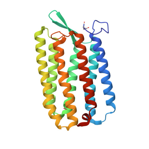
Structures of the archaerhodopsin-3 transporter reveal that disordering of internal water networks underpins receptor sensitization.

Bada Juarez, J.F.Judge, P.J.Adam, S.Axford, D.Vinals, J.Birch, J.Kwan, T.O.C.Hoi, K.K.Yen, H.Y.Vial, A.Milhiet, P.E.Robinson, C.V.Schapiro, I.Moraes, I.Watts, A.

(2021) Nat Commun 12: 629-629

  • DOI: https://doi.org/10.1038/s41467-020-20596-0
  • Primary Citation Related Structures: 
    6GUX, 6S6C

  • PubMed Abstract: 

    Many transmembrane receptors have a desensitized state, in which they are unable to respond to external stimuli. The family of microbial rhodopsin proteins includes one such group of receptors, whose inactive or dark-adapted (DA) state is established in the prolonged absence of light. Here, we present high-resolution crystal structures of the ground (light-adapted) and DA states of Archaerhodopsin-3 (AR3), solved to 1.1 Å and 1.3 Å resolution respectively. We observe significant differences between the two states in the dynamics of water molecules that are coupled via H-bonds to the retinal Schiff Base. Supporting QM/MM calculations reveal how the DA state permits a thermodynamic equilibrium between retinal isomers to be established, and how this same change is prevented in the ground state in the absence of light. We suggest that the different arrangement of internal water networks in AR3 is responsible for the faster photocycle kinetics compared to homologs.


  • Organizational Affiliation
    • Biochemistry Department, Oxford University, South Parks Road, Oxford, OX1 3QU, UK.

Macromolecule Content 

  • Total Structure Weight: 28.66 kDa 
  • Atom Count: 2,259 
  • Modeled Residue Count: 241 
  • Deposited Residue Count: 241 
  • Unique protein chains: 1

Macromolecules

Find similar proteins by:|  3D Structure
Entity ID: 1
MoleculeChains  Sequence LengthOrganismDetailsImage
Archaerhodopsin-3241Halorubrum sodomenseMutation(s): 1 
Membrane Entity: Yes 
UniProt
Find proteins for P96787 (Halorubrum sodomense)
Explore P96787 
Go to UniProtKB:  P96787
Entity Groups
Sequence Clusters30% Identity50% Identity70% Identity90% Identity95% Identity100% Identity
UniProt GroupP96787
Sequence Annotations
Expand
Reference Sequence

Small Molecules

Ligands 7 Unique
IDChains Name / Formula / InChI Key2D Diagram3D Interactions
RET
(Subject of Investigation/LOI)

Query on RET


Download:Ideal Coordinates CCD File
B [auth A]RETINAL
C20 H28 O
NCYCYZXNIZJOKI-OVSJKPMPSA-N
R16

Query on R16


Download:Ideal Coordinates CCD File
C [auth A]
D [auth A]
E [auth A]
F [auth A]
H [auth A]
C [auth A],
D [auth A],
E [auth A],
F [auth A],
H [auth A],
M [auth A]
HEXADECANE
C16 H34
DCAYPVUWAIABOU-UHFFFAOYSA-N
D12

Query on D12


Download:Ideal Coordinates CCD File
G [auth A]DODECANE
C12 H26
SNRUBQQJIBEYMU-UHFFFAOYSA-N
DD9

Query on DD9


Download:Ideal Coordinates CCD File
I [auth A],
J [auth A],
K [auth A],
L [auth A]
nonane
C9 H20
BKIMMITUMNQMOS-UHFFFAOYSA-N
CA

Query on CA


Download:Ideal Coordinates CCD File
N [auth A]CALCIUM ION
Ca
BHPQYMZQTOCNFJ-UHFFFAOYSA-N
CL

Query on CL


Download:Ideal Coordinates CCD File
P [auth A],
Q [auth A],
R [auth A]
CHLORIDE ION
Cl
VEXZGXHMUGYJMC-UHFFFAOYSA-M
NA

Query on NA


Download:Ideal Coordinates CCD File
O [auth A]SODIUM ION
Na
FKNQFGJONOIPTF-UHFFFAOYSA-N
Modified Residues  1 Unique
IDChains TypeFormula2D DiagramParent
PCA
Query on PCA
A
L-PEPTIDE LINKINGC5 H7 N O3GLN

Experimental Data & Validation

Experimental Data

  • Method: X-RAY DIFFRACTION
  • Resolution: 1.07 Å
  • R-Value Free:  0.170 (Depositor), 0.171 (DCC) 
  • R-Value Work:  0.158 (Depositor), 0.158 (DCC) 
  • R-Value Observed: 0.159 (Depositor) 
Space Group: P 21 21 21
Unit Cell:
Length ( Å )Angle ( ˚ )
a = 44.669α = 90
b = 47.357β = 90
c = 104.193γ = 90
Software Package:
Software NamePurpose
REFMACrefinement
DIALSdata reduction
Aimlessdata scaling
PHASERphasing
PDB_EXTRACTdata extraction

Structure Validation

View Full Validation Report


Ligand Structure Quality Assessment 


Entry History 

& Funding Information

Deposition Data


Funding OrganizationLocationGrant Number
Wellcome TrustUnited Kingdom202892/Z/16/Z
Biotechnology and Biological Sciences Research CouncilUnited KingdomBB/N006011/1

Revision History  (Full details and data files)

  • Version 1.0: 2020-07-22
    Type: Initial release
  • Version 1.1: 2021-02-10
    Changes: Database references, Derived calculations
  • Version 1.2: 2024-01-24
    Changes: Advisory, Data collection, Database references, Refinement description
  • Version 1.3: 2024-11-06
    Changes: Structure summary