6S53

Crystal structure of TRIM21 RING domain in complex with an isopeptide-linked Ube2N~ubiquitin conjugate


Experimental Data Snapshot

  • Method: X-RAY DIFFRACTION
  • Resolution: 2.80 Å
  • R-Value Free: 0.249 
  • R-Value Work: 0.209 

wwPDB Validation   3D Report Full Report


Ligand Structure Quality Assessment 


This is version 1.1 of the entry. See complete history


Literature

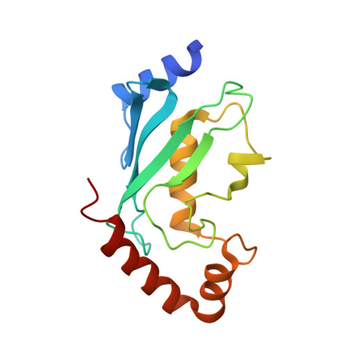
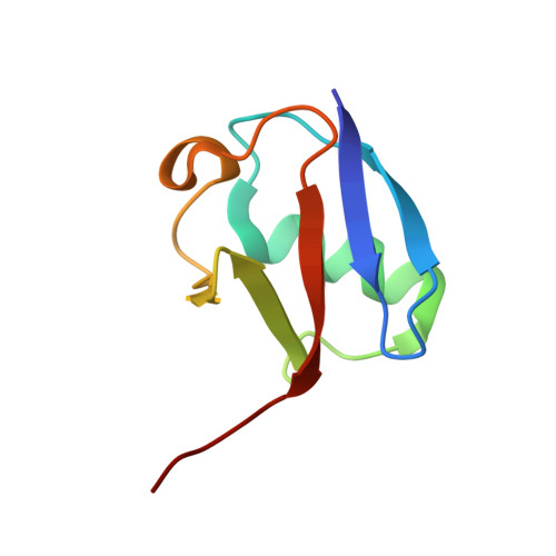
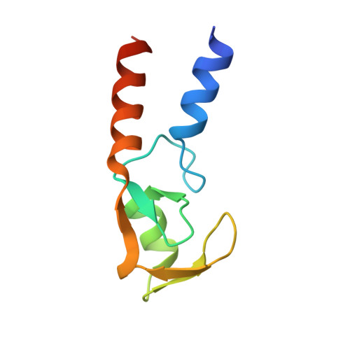
A tri-ionic anchor mechanism drives Ube2N-specific recruitment and K63-chain ubiquitination in TRIM ligases.

Kiss, L.Zeng, J.Dickson, C.F.Mallery, D.L.Yang, J.C.McLaughlin, S.H.Boland, A.Neuhaus, D.James, L.C.

(2019) Nat Commun 10: 4502-4502

  • DOI: https://doi.org/10.1038/s41467-019-12388-y
  • Primary Citation of Related Structures:  
    6S53

  • PubMed Abstract: 
  • The cytosolic antibody receptor TRIM21 possesses unique ubiquitination activity that drives broad-spectrum anti-pathogen targeting and underpins the protein depletion technology Trim-Away. This activity is dependent on formation of self-anchored, K63-linked ubiquitin chains by the heterodimeric E2 enzyme Ube2N/Ube2V2 ...

    The cytosolic antibody receptor TRIM21 possesses unique ubiquitination activity that drives broad-spectrum anti-pathogen targeting and underpins the protein depletion technology Trim-Away. This activity is dependent on formation of self-anchored, K63-linked ubiquitin chains by the heterodimeric E2 enzyme Ube2N/Ube2V2. Here we reveal how TRIM21 facilitates ubiquitin transfer and differentiates this E2 from other closely related enzymes. A tri-ionic motif provides optimally distributed anchor points that allow TRIM21 to wrap an Ube2N~Ub complex around its RING domain, locking the closed conformation and promoting ubiquitin discharge. Mutation of these anchor points inhibits ubiquitination with Ube2N/Ube2V2, viral neutralization and immune signalling. We show that the same mechanism is employed by the anti-HIV restriction factor TRIM5 and identify spatially conserved ionic anchor points in other Ube2N-recruiting RING E3s. The tri-ionic motif is exclusively required for Ube2N but not Ube2D1 activity and provides a generic E2-specific catalysis mechanism for RING E3s.


    Organizational Affiliation

    Medical Research Council Laboratory of Molecular Biology, Cambridge, UK. lcj@mrc-lmb.cam.ac.uk.


Macromolecules
Find similar proteins by:  (by identity cutoff)  |  3D Structure
Entity ID: 1
MoleculeChainsSequence LengthOrganismDetailsImage
Ubiquitin-conjugating enzyme E2 NA [auth E],
C,
G [auth K],
I
152Homo sapiensMutation(s): 0 
Gene Names: UBE2NBLU
EC: 2.3.2.23
UniProt & NIH Common Fund Data Resources
Find proteins for P61088 (Homo sapiens)
Explore P61088 
Go to UniProtKB:  P61088
PHAROS:  P61088
Entity Groups  
Sequence Clusters30% Identity50% Identity70% Identity90% Identity95% Identity100% Identity
UniProt GroupP61088
Protein Feature View
Expand
  • Reference Sequence
Find similar proteins by:  (by identity cutoff)  |  3D Structure
Entity ID: 2
MoleculeChainsSequence LengthOrganismDetailsImage
Polyubiquitin-CB [auth F],
D,
H [auth L],
J
76Homo sapiensMutation(s): 0 
Gene Names: UBC
UniProt & NIH Common Fund Data Resources
Find proteins for P0CG48 (Homo sapiens)
Explore P0CG48 
Go to UniProtKB:  P0CG48
PHAROS:  P0CG48
Entity Groups  
Sequence Clusters30% Identity50% Identity70% Identity90% Identity95% Identity100% Identity
UniProt GroupP0CG48
Protein Feature View
Expand
  • Reference Sequence
Find similar proteins by:  (by identity cutoff)  |  3D Structure
Entity ID: 3
MoleculeChainsSequence LengthOrganismDetailsImage
E3 ubiquitin-protein ligase TRIM21E [auth B],
F [auth A],
K [auth H],
L [auth G]
85Homo sapiensMutation(s): 0 
Gene Names: TRIM21RNF81RO52SSA1
EC: 2.3.2.27
UniProt & NIH Common Fund Data Resources
Find proteins for P19474 (Homo sapiens)
Explore P19474 
Go to UniProtKB:  P19474
PHAROS:  P19474
Entity Groups  
Sequence Clusters30% Identity50% Identity70% Identity90% Identity95% Identity100% Identity
UniProt GroupP19474
Protein Feature View
Expand
  • Reference Sequence
Experimental Data & Validation

Experimental Data

  • Method: X-RAY DIFFRACTION
  • Resolution: 2.80 Å
  • R-Value Free: 0.249 
  • R-Value Work: 0.209 
  • Space Group: P 1
Unit Cell:
Length ( Å )Angle ( ˚ )
a = 49.75α = 89.9
b = 83.31β = 89.05
c = 86.75γ = 88.7
Software Package:
Software NamePurpose
PHENIXrefinement
XDSdata reduction
XDSdata scaling
PHASERphasing

Structure Validation

View Full Validation Report


Ligand Structure Quality Assessment 


Entry History & Funding Information

Deposition Data


Funding OrganizationLocationGrant Number
Medical Research Council (United Kingdom)United KingdomU105181010
Medical Research Council (United Kingdom)United KingdomU105178934
Wellcome TrustUnited KingdomInvestigator Award to Leo C James

Revision History  (Full details and data files)

  • Version 1.0: 2019-09-11
    Type: Initial release
  • Version 1.1: 2019-10-16
    Changes: Data collection, Database references