6S51

The crystal structure of glycogen phosphorylase in complex with 10


Experimental Data Snapshot

  • Method: X-RAY DIFFRACTION
  • Resolution: 2.37 Å
  • R-Value Free: 0.205 
  • R-Value Work: 0.153 
  • R-Value Observed: 0.155 

wwPDB Validation   3D Report Full Report


Ligand Structure Quality Assessment 


This is version 1.0 of the entry. See complete history


Literature

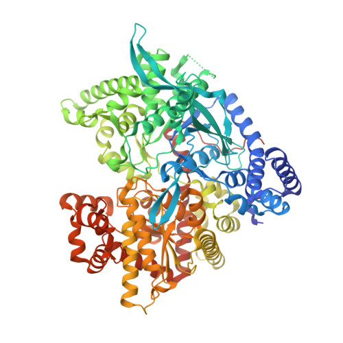
The architecture of hydrogen and sulfur sigma-hole interactions explain differences in the inhibitory potency of C-beta-d-glucopyranosyl thiazoles, imidazoles and an N-beta-d glucopyranosyl tetrazole for human liver glycogen phosphorylase and offer new insights to structure-based design.

Kyriakis, E.Karra, A.G.Papaioannou, O.Solovou, T.Skamnaki, V.T.Liggri, P.G.V.Zographos, S.E.Szennyes, E.Bokor, E.Kun, S.Psarra, A.G.Somsak, L.Leonidas, D.D.

(2020) Bioorg Med Chem 28: 115196-115196

  • DOI: https://doi.org/10.1016/j.bmc.2019.115196
  • Primary Citation of Related Structures:  
    6S4H, 6S4K, 6S4O, 6S4P, 6S4R, 6S51, 6S52

  • PubMed Abstract: 

    C-Glucopyranosyl imidazoles, thiazoles, and an N-glucopyranosyl tetrazole were assessed in vitro and ex vivo for their inhibitory efficiency against isoforms of glycogen phosphorylase (GP; a validated pharmacological target for the development of anti-hyperglycaemic agents). Imidazoles proved to be more potent inhibitors than the corresponding thiazoles or the tetrazole. The most potent derivative has a 2-naphthyl substituent, a K i value of 3.2 µM for hepatic glycogen phosphorylase, displaying also 60% inhibition of GP activity in HepG2 cells, compared to control vehicle treated cells, at 100 μM. X-Ray crystallography studies of the protein - inhibitor complexes revealed the importance of the architecture of inhibitor associated hydrogen bonds or sulfur σ-hole bond interactions to Asn284 OD1, offering new insights to structure-based design efforts. Moreover, while the 2-glucopyranosyl-tetrazole seems to bind differently from the corresponding 1,2,3-triazole compound, the two inhibitors are equipotent.


  • Organizational Affiliation

    Department of Biochemistry and Biotechnology, University of Thessaly, Biopolis, 41500 Larissa, Greece.


Macromolecules
Find similar proteins by:  (by identity cutoff)  |  3D Structure
Entity ID: 1
MoleculeChains Sequence LengthOrganismDetailsImage
Glycogen phosphorylase, muscle form843Oryctolagus cuniculusMutation(s): 0 
EC: 2.4.1.1
UniProt
Find proteins for P00489 (Oryctolagus cuniculus)
Explore P00489 
Go to UniProtKB:  P00489
Entity Groups  
Sequence Clusters30% Identity50% Identity70% Identity90% Identity95% Identity100% Identity
UniProt GroupP00489
Sequence Annotations
Expand
  • Reference Sequence
Small Molecules
Ligands 2 Unique
IDChains Name / Formula / InChI Key2D Diagram3D Interactions
KVQ (Subject of Investigation/LOI)
Query on KVQ

Download Ideal Coordinates CCD File 
B [auth A](2~{R},3~{S},4~{R},5~{R},6~{S})-2-(hydroxymethyl)-6-(2-phenyl-1,3-thiazol-4-yl)oxane-3,4,5-triol
C15 H17 N O5 S
PFYRBHFULSKLRW-RGDJUOJXSA-N
PLP
Query on PLP

Download Ideal Coordinates CCD File 
C [auth A]PYRIDOXAL-5'-PHOSPHATE
C8 H10 N O6 P
NGVDGCNFYWLIFO-UHFFFAOYSA-N
Binding Affinity Annotations 
IDSourceBinding Affinity
KVQ Binding MOAD:  6S51 Ki: 3.26e+5 (nM) from 1 assay(s)
Experimental Data & Validation

Experimental Data

  • Method: X-RAY DIFFRACTION
  • Resolution: 2.37 Å
  • R-Value Free: 0.205 
  • R-Value Work: 0.153 
  • R-Value Observed: 0.155 
  • Space Group: P 43 21 2
Unit Cell:
Length ( Å )Angle ( ˚ )
a = 128.077α = 90
b = 128.077β = 90
c = 115.801γ = 90
Software Package:
Software NamePurpose
REFMACrefinement
CrysalisProdata reduction
Aimlessdata scaling
REFMACphasing

Structure Validation

View Full Validation Report


Ligand Structure Quality Assessment 


Entry History 

Deposition Data

Revision History  (Full details and data files)

  • Version 1.0: 2020-02-19
    Type: Initial release