Millisecond time-resolved serial oscillation crystallography of a blue-light photoreceptor at a synchrotron.
Aumonier, S., Santoni, G., Gotthard, G., von Stetten, D., Leonard, G.A., Royant, A.(2020) IUCrJ 7: 728-736
- PubMed: 32695419 Search on PubMedSearch on PubMed Central
- DOI: https://doi.org/10.1107/S2052252520007411
- Primary Citation Related Structures:
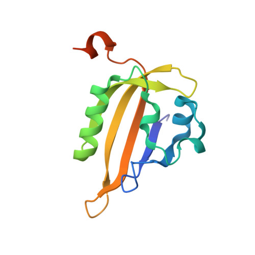
6S45, 6S46 - PubMed Abstract:
The recent development of serial crystallography has popularized time-resolved crystallography as a technique to determine the structure of protein-reaction intermediate states. However, most approaches rely on the availability of thousands to millions of microcrystals. A method is reported here, using monochromatic synchrotron radiation, for the room-temperature collection, processing and merging of X-ray oscillation diffraction data from <100 samples in order to observe the build up of a photoreaction intermediate species. Using this method, we monitored with a time resolution of 63 ms how the population of a blue-light photoreceptor domain in a crystal progressively photoconverts from the dark to the light state. The series of resulting snapshots allows us to visualize in detail the gradual rearrangement of both the protein and chromophore during this process.
- Structural Biology Group, European Synchrotron Radiation Facility, 71 avenue des Martyrs, Grenoble Cedex 9, 38043, France.
Organizational Affiliation: