The structural basis for signal promiscuity in a bacterial chemoreceptor.
Gavira, J.A., Matilla, M.A., Fernandez, M., Krell, T.(2021) FEBS J 288: 2294-2310
- PubMed: 33021055 Search on PubMed
- DOI: https://doi.org/10.1111/febs.15580
- Primary Citation Related Structures:
6S18, 6S1A, 6S33, 6S37, 6S38, 6S3B - PubMed Abstract:
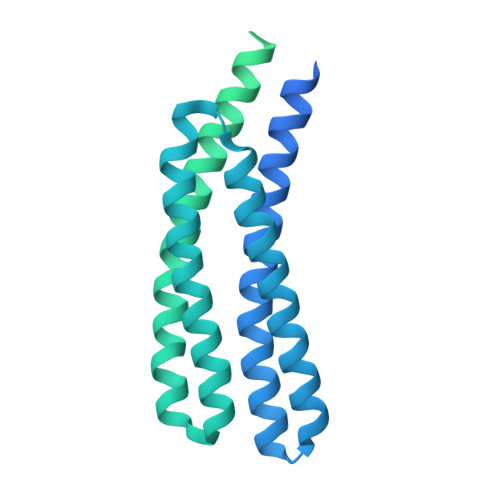
Signalling through chemosensory pathways is typically initiated by the binding of signal molecules to the chemoreceptor ligand binding domain (LBD). The PcaY_PP chemoreceptor from Pseudomonas putida KT2440 is characterized by an unusually broad signal range, and minimal requisites for signal binding are the presence of a C6-membered ring and that of a carboxyl group. Previous studies have shown that only some of the multiple signals recognized by this chemoreceptor are of apparent metabolic value. We report here high-resolution structures of PcaY_PP-LBD in the absence and presence of four cognate chemoeffectors and glycerol. The domain formed a four-helix bundle (4HB), and both ligand binding sites of the dimer were occupied with the high-affinity ligands protocatechuate and quinate, whereas the lower-affinity ligands benzoate and salicylate were present in only one site. Ligand binding was verified by microcalorimetric titration of site-directed mutants revealing important roles of an arginine and number of polar residues that establish an extensive hydrogen bonding network with bound ligands. The comparison of the apo and holo structures did not provide evidence for this receptor employing a transmembrane signalling mechanism that involves piston-like shifts of the final helix. Instead, ligand binding caused rigid-body scissoring movements of both monomers of the dimer. Comparisons with the 4HB domains of the Tar and Tsr chemoreceptors revealed significant structural differences. Importantly, the ligand binding site in PcaY_PP-LBD is approximately 8 Å removed from that of the Tar and Tsr receptors. Data indicate a significant amount of structural and functional diversity among 4HB domains. DATABASES: The coordinates and structure factors have been deposited in the protein data band with the following IDs: 6S1A (apo form), 6S18 (bound glycerol), 6S33 (bound protocatechuate), 6S38 (bound quinate), 6S3B (bound benzoate) and 6S37 (bound salicylate).
- Laboratory of Crystallographic Studies, IACT (CSIC-UGR), Granada, Spain.
Organizational Affiliation: