ParB-type DNA Segregation Proteins Are CTP-Dependent Molecular Switches.
Osorio-Valeriano, M., Altegoer, F., Steinchen, W., Urban, S., Liu, Y., Bange, G., Thanbichler, M.(2019) Cell 179: 1512-1524.e15
- PubMed: 31835030 Search on PubMed
- DOI: https://doi.org/10.1016/j.cell.2019.11.015
- Primary Citation Related Structures:
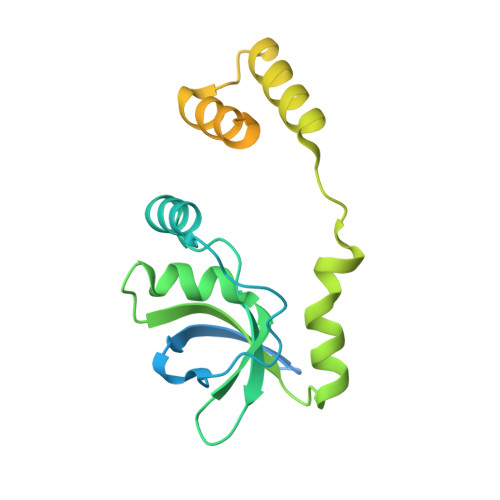
6RYK - PubMed Abstract:
During cell division, newly replicated DNA is actively segregated to the daughter cells. In most bacteria, this process involves the DNA-binding protein ParB, which condenses the centromeric regions of sister DNA molecules into kinetochore-like structures that recruit the DNA partition ATPase ParA and the prokaroytic SMC/condensin complex. Here, we report the crystal structure of a ParB-like protein (PadC) that emerges to tightly bind the ribonucleotide CTP. The CTP-binding pocket of PadC is conserved in ParB and composed of signature motifs known to be essential for ParB function. We find that ParB indeed interacts with CTP and requires nucleotide binding for DNA condensation in vivo. We further show that CTP-binding modulates the affinity of ParB for centromeric parS sites, whereas parS recognition stimulates its CTPase activity. ParB proteins thus emerge as a new class of CTP-dependent molecular switches that act in concert with ATPases and GTPases to control fundamental cellular functions.
- Department of Biology, University of Marburg, 35043 Marburg, Germany; Max Planck Institute for Terrestrial Microbiology, 35043 Marburg, Germany.
Organizational Affiliation: