6RY7 | pdb_00006ry7

Crystal structure of Dfg5 from Chaetomium thermophilum in complex with laminaribiose


Experimental Data Snapshot

  • Method: X-RAY DIFFRACTION
  • Resolution: 1.30 Å
  • R-Value Free: 
    0.119 (Depositor), 0.132 (DCC) 
  • R-Value Work: 
    0.094 (Depositor) 
  • R-Value Observed: 
    0.095 (Depositor) 

wwPDB Validation 3D Report Full Report

Validation slider image for 6RY7

This is version 1.3 of the entry. See complete history

Literature

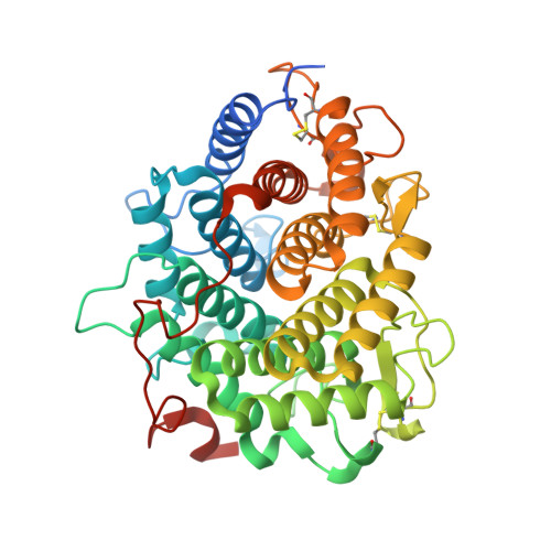
Structural base for the transfer of GPI-anchored glycoproteins into fungal cell walls.

Vogt, M.S.Schmitz, G.F.Varon Silva, D.Mosch, H.U.Essen, L.O.

(2020) Proc Natl Acad Sci U S A 117: 22061-22067

  • DOI: https://doi.org/10.1073/pnas.2010661117
  • Primary Citation Related Structures: 
    6RY0, 6RY1, 6RY2, 6RY5, 6RY6, 6RY7

  • PubMed Abstract: 

    The correct distribution and trafficking of proteins are essential for all organisms. Eukaryotes evolved a sophisticated trafficking system which allows proteins to reach their destination within highly compartmentalized cells. One eukaryotic hallmark is the attachment of a glycosylphosphatidylinositol (GPI) anchor to C-terminal ω-peptides, which are used as a zip code to guide a subset of membrane-anchored proteins through the secretory pathway to the plasma membrane. In fungi, the final destination of many GPI-anchored proteins is their outermost compartment, the cell wall. Enzymes of the Dfg5 subfamily catalyze the essential transfer of GPI-anchored substrates from the plasma membrane to the cell wall and discriminate between plasma membrane-resident GPI-anchored proteins and those transferred to the cell wall (GPI-CWP). We solved the structure of Dfg5 from a filamentous fungus and used in crystallo glycan fragment screening to reassemble the GPI-core glycan in a U-shaped conformation within its binding pocket. The resulting model of the membrane-bound Dfg5•GPI-CWP complex is validated by molecular dynamics (MD) simulations and in vivo mutants in yeast. The latter show that impaired transfer of GPI-CWPs causes distorted cell-wall integrity as indicated by increased chitin levels. The structure of a Dfg5•β1,3-glycoside complex predicts transfer of GPI-CWP toward the nonreducing ends of acceptor glycans in the cell wall. In addition to our molecular model for Dfg5-mediated transglycosylation, we provide a rationale for how GPI-CWPs are specifically sorted toward the cell wall by using GPI-core glycan modifications.


  • Organizational Affiliation
    • Department of Chemistry, Philipps-Universität, D-35032 Marburg, Germany.

Macromolecule Content 

  • Total Structure Weight: 50.08 kDa 
  • Atom Count: 3,866 
  • Modeled Residue Count: 410 
  • Deposited Residue Count: 443 
  • Unique protein chains: 1

Macromolecules

Find similar proteins by:|  3D Structure
Entity ID: 1
MoleculeChains  Sequence LengthOrganismDetailsImage
Mannan endo-1,6-alpha-mannosidase443Thermochaetoides thermophila DSM 1495Mutation(s): 0 
Gene Names: CTHT_0020800
EC: 3.2.1.101
Membrane Entity: Yes 
UniProt
Find proteins for G0S3F2 (Chaetomium thermophilum (strain DSM 1495 / CBS 144.50 / IMI 039719))
Explore G0S3F2 
Go to UniProtKB:  G0S3F2
Entity Groups
Sequence Clusters30% Identity50% Identity70% Identity90% Identity95% Identity100% Identity
UniProt GroupG0S3F2
Sequence Annotations
Expand
Reference Sequence

Oligosaccharides

Help  
Entity ID: 2
MoleculeChains Length2D Diagram GlycosylationD Interactions
beta-D-glucopyranose-(1-3)-beta-D-glucopyranose
B
2N/A
Glycosylation Resources
GlyTouCan: G36535HU
GlyCosmos: G36535HU

Biologically Interesting Molecules (External Reference) 

1 Unique

Experimental Data & Validation

Experimental Data

  • Method: X-RAY DIFFRACTION
  • Resolution: 1.30 Å
  • R-Value Free:  0.119 (Depositor), 0.132 (DCC) 
  • R-Value Work:  0.094 (Depositor) 
  • R-Value Observed: 0.095 (Depositor) 
Space Group: C 1 2 1
Unit Cell:
Length ( Å )Angle ( ˚ )
a = 83.356α = 90
b = 54.879β = 90.45
c = 80.095γ = 90
Software Package:
Software NamePurpose
PHENIXrefinement
XDSdata reduction
Aimlessdata scaling
PHASERphasing

Structure Validation

View Full Validation Report


Entry History 

& Funding Information

Deposition Data


Funding OrganizationLocationGrant Number
German Research FoundationGermanySFB987

Revision History  (Full details and data files)

  • Version 1.0: 2020-08-12
    Type: Initial release
  • Version 1.1: 2020-08-26
    Changes: Database references
  • Version 1.2: 2020-09-16
    Changes: Database references
  • Version 1.3: 2024-10-23
    Changes: Data collection, Database references, Structure summary