X-ray Structures of the Post-fusion 6-Helix Bundle of the Human Syncytins and their Functional Implications.
Ruigrok, K., Vaney, M.C., Buchrieser, J., Baquero, E., Hellert, J., Baron, B., England, P., Schwartz, O., Rey, F.A., Backovic, M.(2019) J Mol Biol 431: 4922-4940
- PubMed: 31711961 Search on PubMedSearch on PubMed Central
- DOI: https://doi.org/10.1016/j.jmb.2019.10.020
- Primary Citation Related Structures:
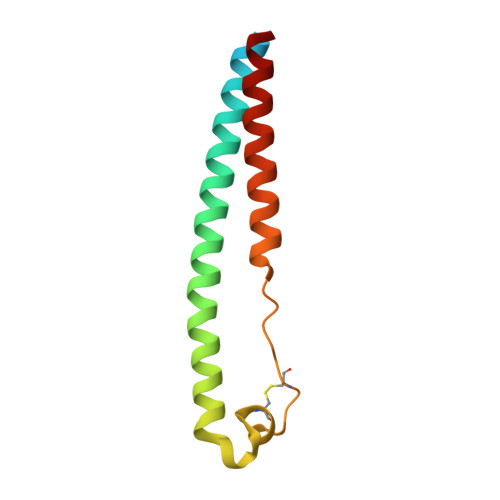
6RX1, 6RX3 - PubMed Abstract:
The retroviral envelope-derived proteins syncytin-1 and syncytin-2 (syn1 and syn2) drive placentation in humans by forming a syncytiotophoblast, a structure allowing for an exchange interface between maternal and fetal blood during pregnancy. Despite their essential role, little is known about the molecular mechanism underlying the syncytins' function. We report here the X-ray structures of the syn1 and syn2 transmembrane subunit ectodomains, featuring a 6-helix bundle (6HB) typical of the post-fusion state of gamma-retrovirus and filovirus fusion proteins. Contrary to the filoviruses, for which the fusion glycoprotein was crystallized both in the post-fusion and in the spring-loaded pre-fusion form, the highly unstable nature of the syncytins' prefusion form has precluded structural studies. We undertook a proline-scanning approach searching for regions in the syn1 6HB central helix that tolerate the introduction of helix-breaker residues and still fold correctly in the pre-fusion form. We found that there is indeed such a region, located two α-helical turns downstream a stutter in the central coiled-coil helix - precisely where the breaks of the spring-loaded helix of the filoviruses map. These mutants were fusion-inactive as they cannot form the 6HB, similar to the "SOSIP" mutant of HIV Env that allowed the high-resolution structural characterization of its labile pre-fusion form. These results now open a new window of opportunity to engineer more stable variants of the elusive pre-fusion trimer of the syncytins and other gamma-retroviruses envelope proteins for structural characterization.
- Unité de Virologie Structurale, Institut Pasteur, Département de Virologie, 28 rue du Dr Roux, 75015 Paris, France; CNRS, UMR3569, Paris, France; Université Paris Descartes, Sorbonne Paris Cité, Paris, France.
Organizational Affiliation: