6RV2

Crystal structure of the human two pore domain potassium ion channel TASK-1 (K2P3.1) in a closed conformation


Experimental Data Snapshot

  • Method: X-RAY DIFFRACTION
  • Resolution: 3.00 Å
  • R-Value Free: 0.257 
  • R-Value Work: 0.253 
  • R-Value Observed: 0.254 

wwPDB Validation   3D Report Full Report


Ligand Structure Quality Assessment 


This is version 1.2 of the entry. See complete history


Literature

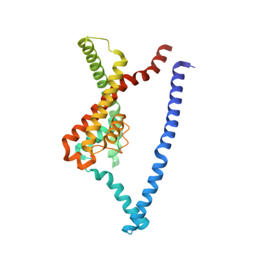
A lower X-gate in TASK channels traps inhibitors within the vestibule.

Rodstrom, K.E.J.Kiper, A.K.Zhang, W.Rinne, S.Pike, A.C.W.Goldstein, M.Conrad, L.J.Delbeck, M.Hahn, M.G.Meier, H.Platzk, M.Quigley, A.Speedman, D.Shrestha, L.Mukhopadhyay, S.M.M.Burgess-Brown, N.A.Tucker, S.J.Muller, T.Decher, N.Carpenter, E.P.

(2020) Nature 582: 443-447

  • DOI: https://doi.org/10.1038/s41586-020-2250-8
  • Primary Citation of Related Structures:  
    6RV2, 6RV3, 6RV4

  • PubMed Abstract: 

    TWIK-related acid-sensitive potassium (TASK) channels-members of the two pore domain potassium (K 2P ) channel family-are found in neurons 1 , cardiomyocytes 2-4 and vascular smooth muscle cells 5 , where they are involved in the regulation of heart rate 6 , pulmonary artery tone 5,7 , sleep/wake cycles 8 and responses to volatile anaesthetics 8-11 . K 2P channels regulate the resting membrane potential, providing background K + currents controlled by numerous physiological stimuli 12-15 . Unlike other K 2P channels, TASK channels are able to bind inhibitors with high affinity, exceptional selectivity and very slow compound washout rates. As such, these channels are attractive drug targets, and TASK-1 inhibitors are currently in clinical trials for obstructive sleep apnoea and atrial fibrillation 16 . In general, potassium channels have an intramembrane vestibule with a selectivity filter situated above and a gate with four parallel helices located below; however, the K 2P channels studied so far all lack a lower gate. Here we present the X-ray crystal structure of TASK-1, and show that it contains a lower gate-which we designate as an 'X-gate'-created by interaction of the two crossed C-terminal M4 transmembrane helices at the vestibule entrance. This structure is formed by six residues ( 243 VLRFMT 248 ) that are essential for responses to volatile anaesthetics 10 , neurotransmitters 13 and G-protein-coupled receptors 13 . Mutations within the X-gate and the surrounding regions markedly affect both the channel-open probability and the activation of the channel by anaesthetics. Structures of TASK-1 bound to two high-affinity inhibitors show that both compounds bind below the selectivity filter and are trapped in the vestibule by the X-gate, which explains their exceptionally low washout rates. The presence of the X-gate in TASK channels explains many aspects of their physiological and pharmacological behaviour, which will be beneficial for the future development and optimization of TASK modulators for the treatment of heart, lung and sleep disorders.


  • Organizational Affiliation

    Structural Genomics Consortium, University of Oxford, Oxford, UK.


Macromolecules
Find similar proteins by:  (by identity cutoff)  |  3D Structure
Entity ID: 1
MoleculeChains Sequence LengthOrganismDetailsImage
Potassium channel subfamily K member 3
A, B, C, D
264Homo sapiensMutation(s): 0 
Gene Names: KCNK3TASKTASK1
Membrane Entity: Yes 
UniProt & NIH Common Fund Data Resources
Find proteins for O14649 (Homo sapiens)
Explore O14649 
Go to UniProtKB:  O14649
PHAROS:  O14649
GTEx:  ENSG00000171303 
Entity Groups  
Sequence Clusters30% Identity50% Identity70% Identity90% Identity95% Identity100% Identity
UniProt GroupO14649
Sequence Annotations
Expand
  • Reference Sequence
Small Molecules
Ligands 4 Unique
IDChains Name / Formula / InChI Key2D Diagram3D Interactions
PC1
Query on PC1

Download Ideal Coordinates CCD File 
K [auth A],
P [auth B],
W [auth D],
Z [auth D]
1,2-DIACYL-SN-GLYCERO-3-PHOSPHOCHOLINE
C44 H88 N O8 P
NRJAVPSFFCBXDT-HUESYALOSA-N
Y01
Query on Y01

Download Ideal Coordinates CCD File 
H [auth A]
I [auth A]
M [auth A]
N [auth B]
U [auth C]
H [auth A],
I [auth A],
M [auth A],
N [auth B],
U [auth C],
X [auth D],
Y [auth D]
CHOLESTEROL HEMISUCCINATE
C31 H50 O4
WLNARFZDISHUGS-MIXBDBMTSA-N
DMU
Query on DMU

Download Ideal Coordinates CCD File 
J [auth A],
O [auth B],
V [auth C]
DECYL-BETA-D-MALTOPYRANOSIDE
C22 H42 O11
WOQQAWHSKSSAGF-WXFJLFHKSA-N
K
Query on K

Download Ideal Coordinates CCD File 
E [auth A]
F [auth A]
G [auth A]
L [auth A]
Q [auth C]
E [auth A],
F [auth A],
G [auth A],
L [auth A],
Q [auth C],
R [auth C],
S [auth C],
T [auth C]
POTASSIUM ION
K
NPYPAHLBTDXSSS-UHFFFAOYSA-N
Experimental Data & Validation

Experimental Data

  • Method: X-RAY DIFFRACTION
  • Resolution: 3.00 Å
  • R-Value Free: 0.257 
  • R-Value Work: 0.253 
  • R-Value Observed: 0.254 
  • Space Group: P 2 21 21
Unit Cell:
Length ( Å )Angle ( ˚ )
a = 45.14α = 90
b = 204.79β = 90
c = 238.34γ = 90
Software Package:
Software NamePurpose
BUSTERrefinement
XDSdata reduction
XSCALEdata scaling
STARANISOdata scaling
PHASERphasing

Structure Validation

View Full Validation Report


Ligand Structure Quality Assessment 


Entry History & Funding Information

Deposition Data


Funding OrganizationLocationGrant Number
Wellcome TrustUnited Kingdom106169/Z/14/Z
European CommissionUnited Kingdom115766
Biotechnology and Biological Sciences Research CouncilUnited KingdomBB/N009274/1

Revision History  (Full details and data files)

  • Version 1.0: 2019-08-07
    Type: Initial release
  • Version 1.1: 2020-07-22
    Changes: Database references
  • Version 1.2: 2024-01-24
    Changes: Data collection, Database references, Derived calculations, Refinement description